
Pediatric Ophthalmology and Strabismus — MCQs

On this page
Onset of stereopsis occurs at the age of:
Crossed eye fixation is positive in -
Which of the following statements about the treatment of cataract in children is false?
Which of the following statements about divergent squint is true?
Which ocular manifestation is considered a hallmark feature of Goldenhar syndrome?
In the context of ophthalmology, what does the acronym BSGT refer to?
Which muscle paralysis causes right esotropia?
Esotropia is common in
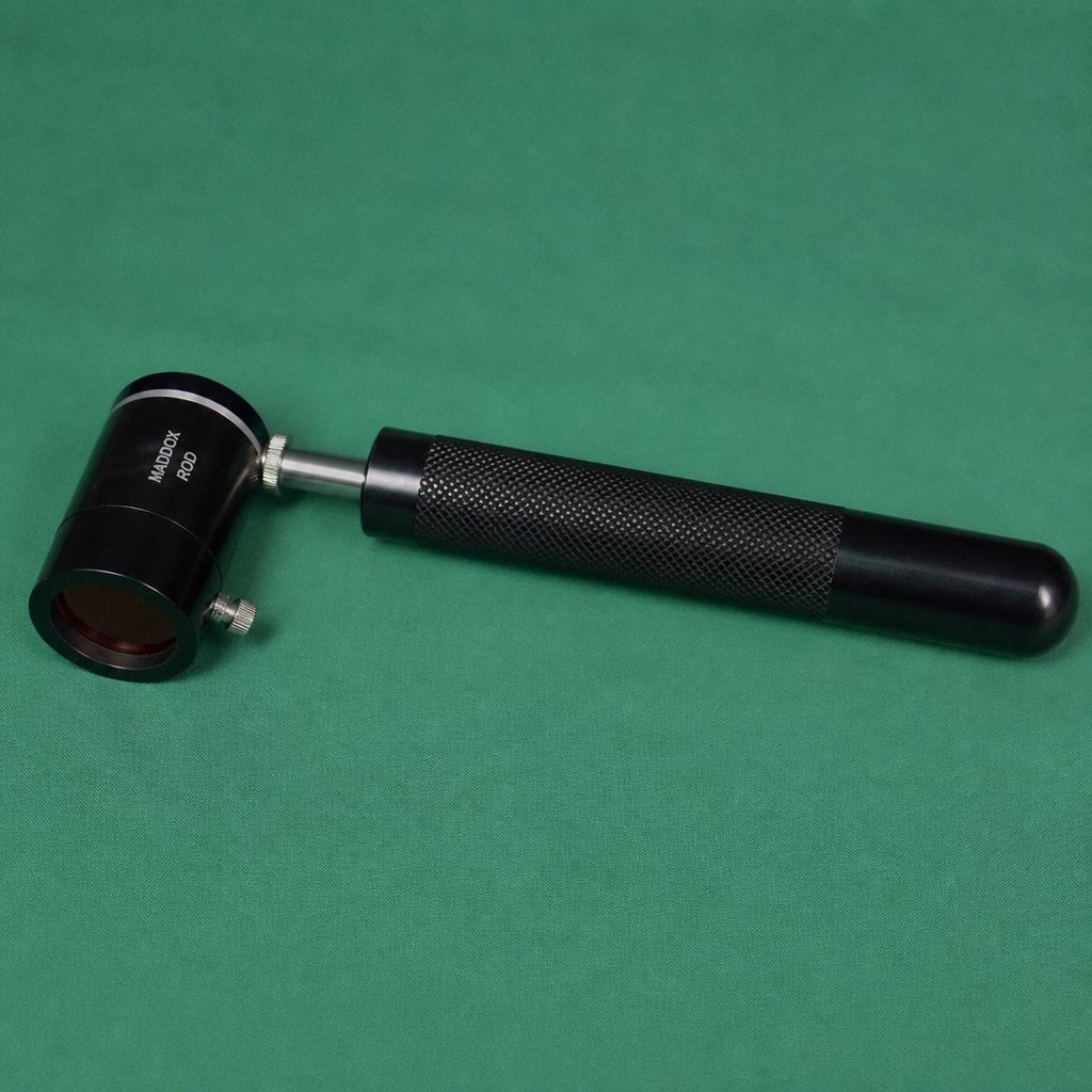
Identify the ophthalmic instrument used for measuring heterophoria and heterotropia.
A 65-year-old male with a history of hypertension and diabetes presents to the OPD with complaints of diplopia and squint. On examination, the secondary deviation is more than the primary deviation. Which of the following is the most likely diagnosis?
Practice by Chapter
Amblyopia
Practice Questions
Esotropia
Practice Questions
Exotropia
Practice Questions
Vertical Deviations
Practice Questions
Special Forms of Strabismus
Practice Questions
Nystagmus in Children
Practice Questions
Pediatric Cataract
Practice Questions
Retinopathy of Prematurity
Practice Questions
Pediatric Glaucoma
Practice Questions
Pediatric Neuro-ophthalmology
Practice Questions
Genetic Eye Diseases in Children
Practice Questions
Pediatric Ocular Trauma
Practice Questions
Want unlimited practice?
Get full access to all questions, explanations, and performance tracking.
Scan to download app