
Orbital Diseases — MCQs

On this page
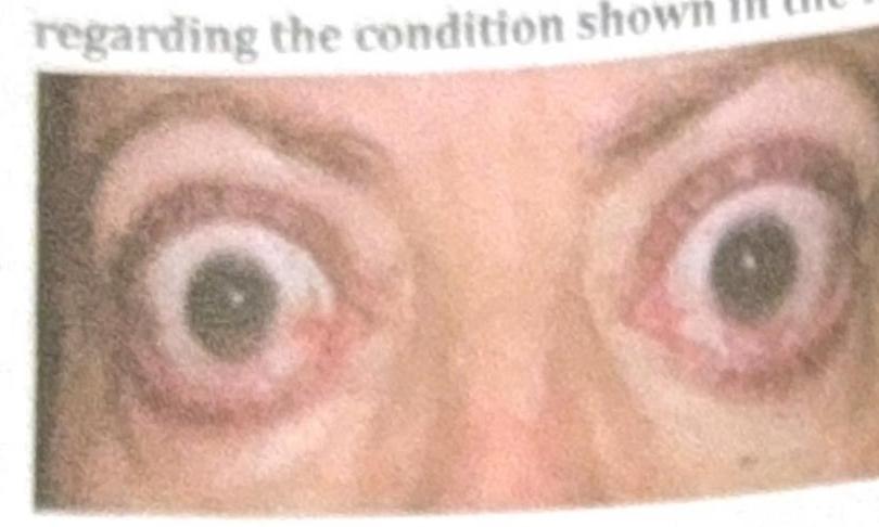
Which of the following statements about thyroid eye disease is false?
The most common benign tumour of the orbit is
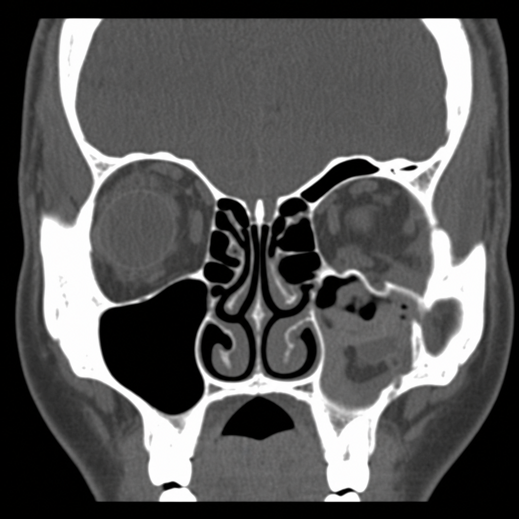
A patient presents to the emergency department after sustaining blunt trauma to the left eye. Clinical examination reveals enophthalmos, diplopia on upward gaze, and periorbital ecchymosis. The CT scan image is shown below. What is your diagnosis?
What is the diagnosis for a patient with unilateral proptosis with bilateral 6th nerve palsy with chemosis and euthyroid status?
The most common cause of proptosis in adults:-
All are causes of proptosis except:
A 30-year-old female presents with proptosis, pain, and vision loss in one eye. An MRI reveals an orbital mass. What is the most likely diagnosis?
A 5-year-old child presents with painless, progressive proptosis of the right eye. MRI reveals a mass in the right orbit. Which structure is most likely involved?
The globe is displaced to which side in lacrimal gland tumour?
Enophthalmos is seen in ?
Practice by Chapter
Orbital Anatomy
Practice Questions
Orbital Imaging Techniques
Practice Questions
Orbital Inflammations
Practice Questions
Orbital Infections
Practice Questions
Orbital Tumors: Primary
Practice Questions
Orbital Tumors: Secondary
Practice Questions
Vascular Lesions of Orbit
Practice Questions
Thyroid Orbitopathy
Practice Questions
Orbital Trauma
Practice Questions
Congenital Orbital Anomalies
Practice Questions
Orbital Surgery Techniques
Practice Questions
Enucleation and Exenteration
Practice Questions
Want unlimited practice?
Get full access to all questions, explanations, and performance tracking.
Scan to download app