
Orbital Diseases — MCQs

On this page
A patient has bilateral proptosis with restricted movements of the eye. What is the most common cause of such condition?
What is the cause of pulsating proptosis?
Exophthalmos is not typically present in which of the following conditions?
Axial proptosis is associated with which of the following?
Which one of the following is the least likely cause of a patient's symptoms of retrobulbar swelling and diplopia?
Which clinical feature differentiates orbital apex syndrome from superior orbital fissure syndrome?
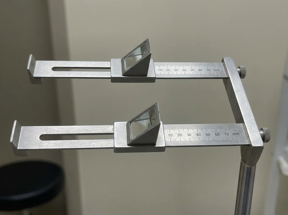
Which instrument is shown in the given image?
A 25-year-old diabetic patient presents with a hectic fever, bilateral proptosis, and a history of facial injury. What is the most likely diagnosis?
The 24-12-6 rule is applied for which type of surgery?
A tumor has the following characteristics: retrobulbar location within the muscle cone, well-defined capsule, presents with slowly progressive proptosis, is easily resectable, and occurs most commonly in the 2nd to 4th decade. What is the most likely diagnosis?
Practice by Chapter
Orbital Anatomy
Practice Questions
Orbital Imaging Techniques
Practice Questions
Orbital Inflammations
Practice Questions
Orbital Infections
Practice Questions
Orbital Tumors: Primary
Practice Questions
Orbital Tumors: Secondary
Practice Questions
Vascular Lesions of Orbit
Practice Questions
Thyroid Orbitopathy
Practice Questions
Orbital Trauma
Practice Questions
Congenital Orbital Anomalies
Practice Questions
Orbital Surgery Techniques
Practice Questions
Enucleation and Exenteration
Practice Questions
Want unlimited practice?
Get full access to all questions, explanations, and performance tracking.
Scan to download app