Which of the following is the first visual field defect in open-angle glaucoma?
A patient complains of an inability to read a newspaper, particularly in bright sunlight. What is the most likely diagnosis?
According to WHO ICD-11 classification, visual impairment in the better eye (with best correction) begins at a visual acuity worse than:
A 12-year-old boy is admitted to the emergency department with signs of meningitis. To determine the specific type of meningitis, it is necessary to aspirate cerebrospinal fluid with a lumbar puncture for laboratory examination. However, before performing a lumbar puncture, it must be established that the cerebrospinal fluid pressure is not elevated. What condition in the eye would indicate that cerebrospinal fluid pressure is too elevated for a lumbar puncture to be performed?
Under Vision 2020, to check visual acuity, a teacher will refer a school child to
Which of the following best predicts the need for lumbar puncture in a patient with syphilis?
Maximum correction of myopia can be done by?
1mm change in axial length of the eyeball would change the refracting power of the eye by?
A person with a visual acuity of 6/60 in the right eye and 3/60 in the left eye would be categorized into which type of blindness?
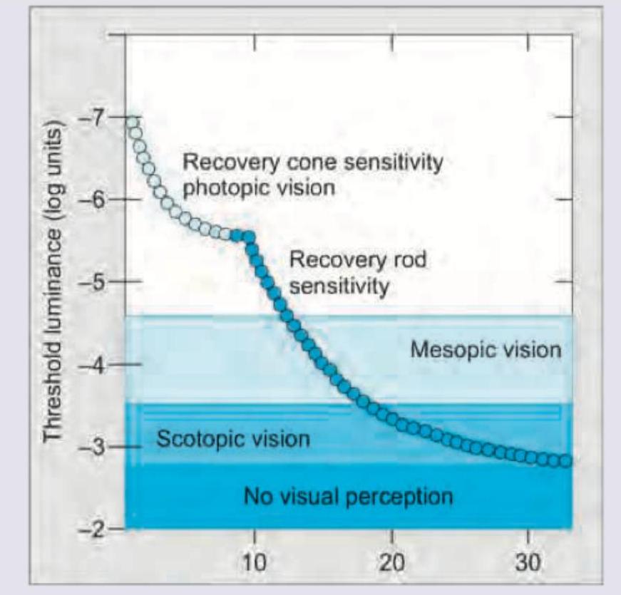
The image given below shows:

Explanation: ***Paracentral scotoma*** - This is the **earliest visual field defect** detected in open-angle glaucoma, typically appearing in the **Bjerrum area** (10-20° from fixation). - Most commonly occurs as a **superior or inferior arcuate scotoma** in the nasal field. - Results from damage to the **retinal nerve fiber layer** around the **optic disc**, which is particularly vulnerable to elevated intraocular pressure. - These scotomas respect the **horizontal raphe** and follow the arcuate nerve fiber bundle pattern. *Ring scotoma* - A **ring scotoma** (Bjerrum scotoma) typically occurs later in the progression of glaucoma, when superior and inferior arcuate defects coalesce to form a ring-like pattern. - This represents **advanced glaucomatous damage** and is not an early finding. *Bitemporal hemianopia* - This visual field defect is characteristic of **optic chiasm compression**, commonly due to a **pituitary tumor** or other suprasellar lesions. - It is **not associated with glaucoma**, which causes damage to the optic nerve fibers within the eye, not at the chiasm. *Tunnel vision* - **Tunnel vision** represents severe, **end-stage glaucoma**, where only a small central island of vision remains. - It indicates extensive loss of peripheral visual field and is a late finding, not an early one.
Explanation: ***Posterior subcapsular cataract*** - This type of cataract causes significant **glare** and **photophobia**, making it difficult to read in bright light due to opacities located at the **posterior lens capsule**. - The patient experiences worsening vision in **bright light** conditions because the constricted pupil directs more light through the **central posterior opacity**, which lies directly in the visual axis. *Nuclear cataract* - Patients with **nuclear cataracts** typically experience **myopic shift** and improved near vision (second sight) due to increased refractive power of the lens. - Vision is usually worse in **dim light** conditions because of pupillary dilation, which allows more light to pass through the central opacity. *Cortical cataract* - Characterized by **spoke-like opacities** that start in the periphery and extend inward. - While it can cause glare, vision often remains good until the opacities encroach upon the **visual axis**, and it doesn't specifically cause worsening vision in bright light to the same degree as PSC. *Congenital cataract* - Present at birth or shortly after, and symptoms depend on the density and location of the opacity. - While it affects vision, the specific complaint of difficulty reading in bright sunlight is not a typical distinguishing feature of **congenital cataracts**.
Explanation: ***6/12*** - According to the **WHO ICD-11 classification**, visual impairment begins when visual acuity is **less than 6/12** in the better eye with best correction. - This threshold marks the beginning of **mild visual impairment** (visual acuity < 6/12 to ≥ 6/18). - Visual acuity of 6/12 or better is considered **normal vision** without significant impairment. *6/24* - Visual acuity of **6/24** falls within the **moderate visual impairment** category (< 6/18 to ≥ 6/60). - This represents established visual impairment but is not the threshold where impairment begins. *6/36* - Visual acuity of **6/36** also falls within the **moderate visual impairment** range. - This indicates more significant vision loss than the threshold that defines the beginning of visual impairment. *6/60* - Visual acuity of **6/60 or worse** (< 6/60 to ≥ 3/60) is classified as **severe visual impairment**. - Visual acuity worse than 3/60 is classified as **blindness**. - This represents much more severe vision loss than the initial threshold for visual impairment.
Explanation: ***Papilledema*** - **Papilledema** is **swelling of the optic disc** due to increased intracranial pressure (ICP), which can be visualized during an **ophthalmoscopic examination**. - Performing a **lumbar puncture (LP)** in the presence of papilledema can lead to **brain herniation** due to a sudden drop in pressure below the spinal cord, creating a pressure gradient that forces brain tissue downward. - This is the **primary ocular contraindication** to LP and must be assessed before the procedure. *Retinal hemorrhages at the fovea* - **Retinal hemorrhages** at the fovea are not a direct sign of **increased intracranial pressure (ICP)** in the same way papilledema is. - While certain conditions causing elevated ICP can lead to retinal hemorrhages (e.g., severe hypertension, Terson syndrome), they are not the primary or most reliable indicator for contraindicating an **LP** compared to papilledema. - Retinal hemorrhages can occur from various causes including diabetic retinopathy, retinal vein occlusion, or trauma. *Obvious opacity of the lens* - An **obvious opacity of the lens** refers to a **cataract**. - **Cataracts** are lens opacities that impair vision and are typically associated with aging, trauma, or systemic diseases like diabetes, but not directly with **elevated CSF pressure** or as a contraindication for **LP**. *Separation of the pars optica retinae anterior to the ora serrata* - The **pars optica retinae** refers to the photosensitive posterior part of the retina, and the **ora serrata** is its anterior boundary. - **Separation** in this area might suggest a **retinal detachment** or other structural retinal issue, which is not an indicator of **elevated intracranial pressure (ICP)** and would not contraindicate a **lumbar puncture**.
Explanation: ***Vision centre*** - Under Vision 2020 initiatives, a **Vision Centre** serves as the primary point of contact for basic ophthalmic services, including **visual acuity screening** and referral. - These centers are designed to be accessible in local communities, allowing teachers and other local caregivers to refer school children for initial checks and appropriate management. *Centre for excellence* - A **Centre for Excellence** typically refers to a highly specialized institution with advanced diagnostic and treatment capabilities, research facilities, and complex surgical procedures, which is **beyond the scope** of basic visual acuity checking and initial referral. - Such centers handle more **complex or rare conditions** and are not the first point of contact for routine school-based screening. *Training centre* - A **Training Centre** is primarily dedicated to educating and skilling healthcare professionals, not to providing direct patient care or screening services to the general public. - While essential for developing skilled personnel, it is **not the appropriate facility** for a teacher to refer a child for a visual acuity check. *Service centre* - The term **Service Centre** is too broad and can refer to various types of facilities that provide any kind of service, but it does not specifically denote a healthcare facility for ophthalmic care under the Vision 2020 program. - It lacks the **specific medical context** and structured role established for vision screening.
Explanation: ***Neurological or ophthalmic signs*** - The presence of **neurological symptoms** (e.g., headache, confusion, motor deficits) or **ophthalmic abnormalities** (e.g., vision changes, uveitis) in a patient with syphilis strongly indicates central nervous system (CNS) involvement, necessitating a **lumbar puncture** to diagnose **neurosyphilis**. - These signs suggest the spirochete has invaded the CNS or eye, making CSF examination critical for confirming involvement and guiding treatment. *Presence of genital ulcers* - Genital ulcers are characteristic of **primary syphilis** and do not directly predict CNS involvement or the need for a lumbar puncture. - While they indicate active infection, the infection may not have progressed to the CNS at this stage. *History of prior treatment* - A history of prior treatment for syphilis, even if adequately treated, does not inherently indicate a need for lumbar puncture unless there is evidence of **treatment failure** or **neurosyphilis symptoms**. - Retreatment is usually guided by serological response or clinical symptoms, not just a history of prior treatment. *Positive VDRL test* - A positive **Venereal Disease Research Laboratory (VDRL)** test confirms active syphilis infection and is used for monitoring treatment response. - However, it does not specifically indicate CNS involvement and is not a sole predictor for performing a lumbar puncture without accompanying neurological or ophthalmological signs.
Explanation: ***LASIK*** - **LASIK (Laser-Assisted In Situ Keratomileusis)** allows for significant correction of high myopia by reshaping the cornea with an excimer laser. - It involves creating a **corneal flap** and then ablating tissue underneath, offering precise and stable vision correction for a wide range of refractive errors. - Among the given corneal refractive procedures, LASIK can correct myopia up to **-10 to -12 D**. *Radial keratotomy* - **Radial keratotomy (RK)** involves making radial incisions in the cornea to flatten it, primarily used for low to moderate myopia (up to -3 to -4 D). - It has a higher risk of **unpredictable outcomes**, induced astigmatism, and glare compared to modern laser procedures. *Photorefractive keratectomy* - **Photorefractive keratectomy (PRK)** involves direct ablation of the corneal surface without creating a flap, which is suitable for moderate myopia (up to -8 to -10 D). - While effective, PRK typically has a **longer recovery period** and more post-operative pain than LASIK. *Orthokeratology* - **Orthokeratology (Ortho-K)** uses specially designed rigid contact lenses worn overnight to temporarily reshape the cornea and correct myopia. - The effect is **temporary**, requiring continuous lens wear to maintain vision correction, and is generally limited to low to moderate myopia (up to -4 to -6 D).
Explanation: ***3D*** - A 1mm change in the **axial length** of the eyeball leads to an approximate **3 diopter (D) change** in the refractive power of the eye. - This relationship is crucial for understanding **refractive errors** like myopia (if the eyeball is too long) or hyperopia (if it's too short). *1D* - A 1D change in refractive power corresponds to a much larger change in the **focal length** of the eye, not typically 1mm in axial length. - This value is too small to reflect the significant impact of a 1mm axial length alteration on the eye's focusing ability. *2D* - While a direct relationship exists, 2D is an **underestimation** of the actual refractive change caused by a 1mm alteration in axial length. - This value would imply a less sensitive optical system than the human eye. *4D* - A 4D change would represent an **overestimation** of the refractive power change for a 1mm alteration in axial length. - Such a high value is generally seen with more substantial anatomical variations or surgical interventions.
Explanation: ***Moderate visual impairment*** - According to the **WHO International Classification of Diseases (ICD-11)**, moderate visual impairment is defined as visual acuity **< 6/18 to ≥ 6/60** in the better eye with best possible correction. - This patient's better eye (right eye) has a visual acuity of **6/60**, which falls at the **upper limit** of the moderate visual impairment category. - This is the standard classification used in Indian medical examinations and follows WHO guidelines. *Legal blindness* - **Legal blindness** is a **US administrative/legal term**, not a WHO classification category. It is defined as visual acuity **< 6/60 (or 20/200)** in the better eye, or visual field < 20 degrees. - Since this patient has exactly **6/60** (not less than 6/60), they do **not** meet the strict criteria for legal blindness. - This term is less commonly used in Indian medical literature, where WHO classifications are standard. *Low vision* - **Low vision** is a broad umbrella term that includes all categories of visual impairment from mild to severe, but it is not a specific classification category. - While this patient does have low vision, the more specific and appropriate classification is moderate visual impairment. *Normal vision* - **Normal vision** is defined as visual acuity of **6/6 to 6/12** in the better eye. - This patient's visual acuity of **6/60** represents significant visual impairment, far below the normal range.
Explanation: ***Dark adaptation curve*** - The image displays the change in **threshold luminance** over time, specifically showing two distinct phases of recovery of sensitivity: an initial rapid phase (cones) and a later, slower, more sensitive phase (rods). - This bimodal curve is characteristic of the **dark adaptation process**, where the eye adjusts from bright to dim light, increasing its sensitivity to light stimuli. *Visual evoked response* - A **visual evoked response (VER)** measures the electrical signals generated in the brain in response to visual stimuli. - VER graphs typically show amplitude and latency of brain activity, not a curve of threshold luminance over time. *Contrast sensitivity plot* - A **contrast sensitivity plot** illustrates the ability to distinguish between different levels of contrast at various spatial frequencies. - This is usually depicted as a curve showing contrast sensitivity as a function of spatial frequency, which is different from the time-dependent threshold luminance shown. *Electroretinography curve* - **Electroretinography (ERG)** measures the electrical responses of various retinal cell types to light stimuli. - An ERG curve typically shows a characteristic waveform with specific a-wave and b-wave components, representing photoreceptor and bipolar cell activity, which is not what is presented in the image.
Get full access to all questions, explanations, and performance tracking.
Start For Free