
Optics and Refraction — MCQs

On this page
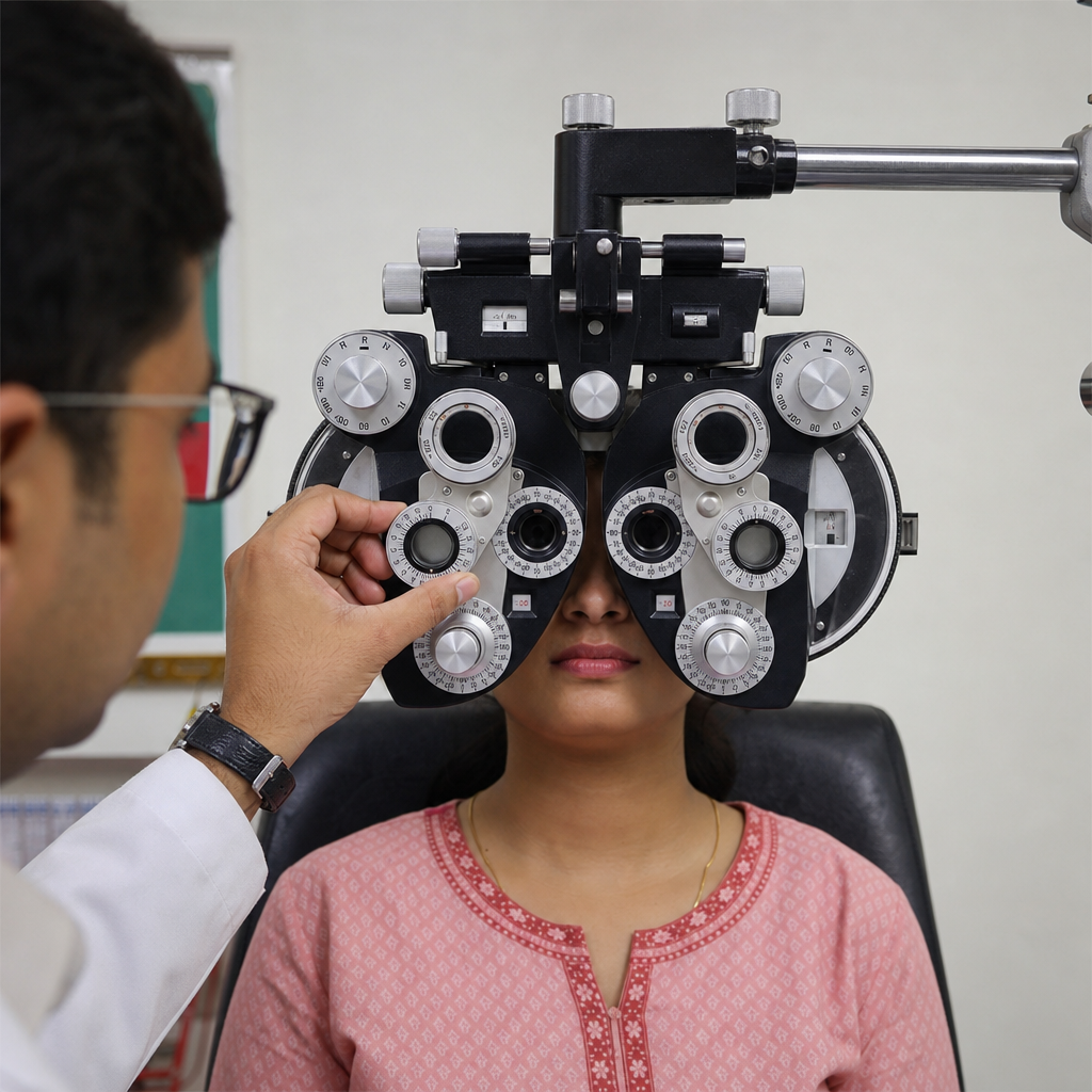
The optical procedure being done is used for:
When water enters the eyes, blurring of vision is due to which of the following?
What is the anterior focal length of the schematic eye?
Which condition is characterized by tubular vision?
What is Sturm's Conoid?
What is the most important factor determining the convergence of light rays on the retina?
What is the magnification obtained with a direct ophthalmoscope?
What is the power of a lens with a focal length of 0.75 meters?
What is the primary treatment for presbyopia?
Which of the following is the most important factor for refractive errors?
Practice by Chapter
Physical Optics
Practice Questions
Geometric Optics
Practice Questions
Optical System of Eye
Practice Questions
Visual Acuity and Contrast Sensitivity
Practice Questions
Refractive Errors
Practice Questions
Accommodation and Presbyopia
Practice Questions
Optical Instruments
Practice Questions
Lenses and Prisms
Practice Questions
Retinoscopy
Practice Questions
Subjective Refraction
Practice Questions
Contact Lens Optics
Practice Questions
Wavefront Technology
Practice Questions
Want unlimited practice?
Get full access to all questions, explanations, and performance tracking.
Scan to download app