
Optics and Refraction — MCQs

On this page
Which of the following is NOT a part of accommodation?
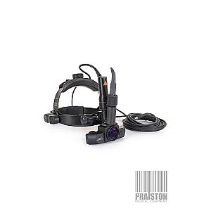
What is the power of the lens used to view the retina with this instrument?
Minus cylinder lenses are prescribed because of what reason?
On performing retinoscopy, in which of the following conditions does the movement of the red reflex not occur with the movement of the retinoscope?
What is the most accepted modality of treatment for 2D myopia in a 13-year-old girl?
The prism produces displacement of the objects seen through it towards the:
True regarding retinoscopy are all of the following except?
If the refractive power of the unaccommodated eye of an emmetropic woman is 60 diopters (D), what is the approximate axial length of her eye?
The total refractive power of the eye is normally 59 diopters. Two-thirds of this power is contributed by which ocular structure?
What is the angle subtended by the topmost letter in the Snellen's chart at the nodal point of the eye when the person is viewing it from 6 meters?
Practice by Chapter
Physical Optics
Practice Questions
Geometric Optics
Practice Questions
Optical System of Eye
Practice Questions
Visual Acuity and Contrast Sensitivity
Practice Questions
Refractive Errors
Practice Questions
Accommodation and Presbyopia
Practice Questions
Optical Instruments
Practice Questions
Lenses and Prisms
Practice Questions
Retinoscopy
Practice Questions
Subjective Refraction
Practice Questions
Contact Lens Optics
Practice Questions
Wavefront Technology
Practice Questions
Want unlimited practice?
Get full access to all questions, explanations, and performance tracking.
Scan to download app