
Optics and Refraction — MCQs

On this page
What is the most common type of refractive error?
For a near point of 20 cm, a hypermetropic patient with +4D refractive error has to exercise what amount of accommodation?
In retinoscopy performed with a plane mirror at a distance of 1 meter, if the retinal glow moves in the opposite direction to the mirror's movement, what refractive error does the patient have?
Which of the following is not a feature of pathological myopia?
A patient complains of distorted vision after wearing spectacles, which worsens progressively in both meridians. Which of the following is NOT true about this condition?
A patient with open-angle glaucoma and 7D of myopia complains of blurring of vision after receiving pilocarpine. What is the reason for this blurring?
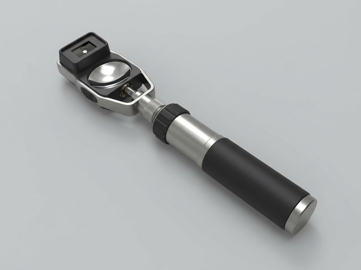
Which of the following procedures or tests is NOT performed using this instrument?
Which statement is true about Jackson's Cross Cylinder test?
What is keratometry used for?
What are the refractive indices of the nucleus and cortex of the lens, respectively?
Practice by Chapter
Physical Optics
Practice Questions
Geometric Optics
Practice Questions
Optical System of Eye
Practice Questions
Visual Acuity and Contrast Sensitivity
Practice Questions
Refractive Errors
Practice Questions
Accommodation and Presbyopia
Practice Questions
Optical Instruments
Practice Questions
Lenses and Prisms
Practice Questions
Retinoscopy
Practice Questions
Subjective Refraction
Practice Questions
Contact Lens Optics
Practice Questions
Wavefront Technology
Practice Questions
Want unlimited practice?
Get full access to all questions, explanations, and performance tracking.
Scan to download app