The chart shown in the image is:

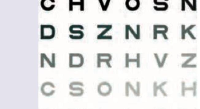
The following alphabet of Snellen's chart will subtend an angle of how many minutes at nodal point of the eye?

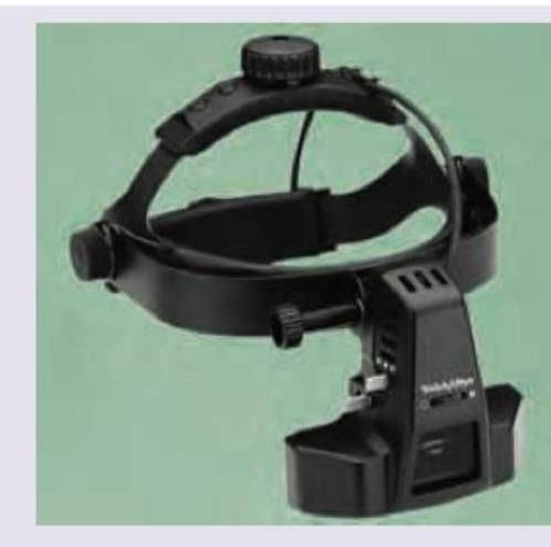
What is the power of lens attached to this instrument to visualize the entire retina?

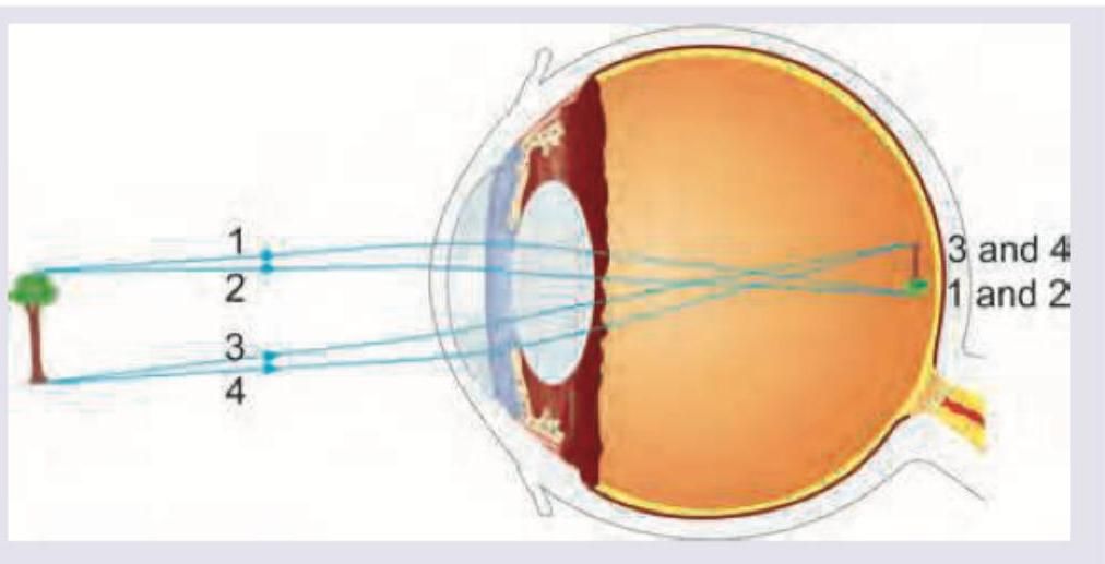
Identify the refractive error shown in the image:

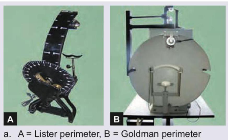
Identify the instrument shown:

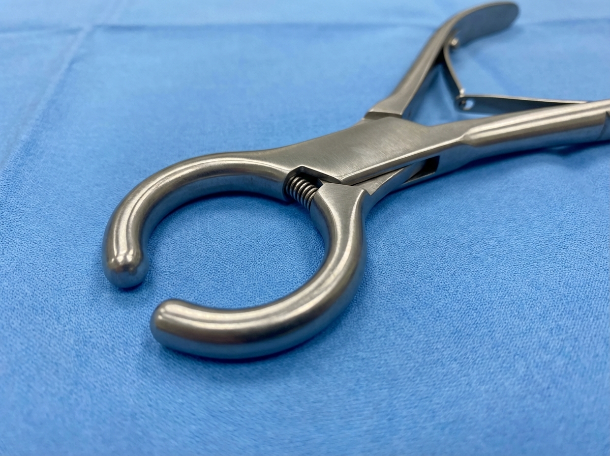
Identify the instrument? (NEET Pattern 2019)

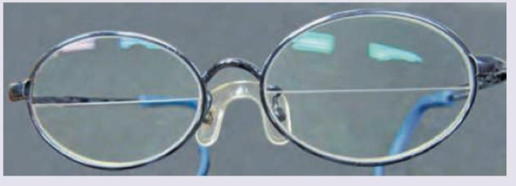
The following spectacle is used in? (AIIMS Nov 2018)

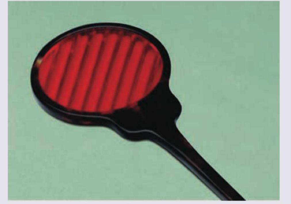
Identify the instrument shown below:

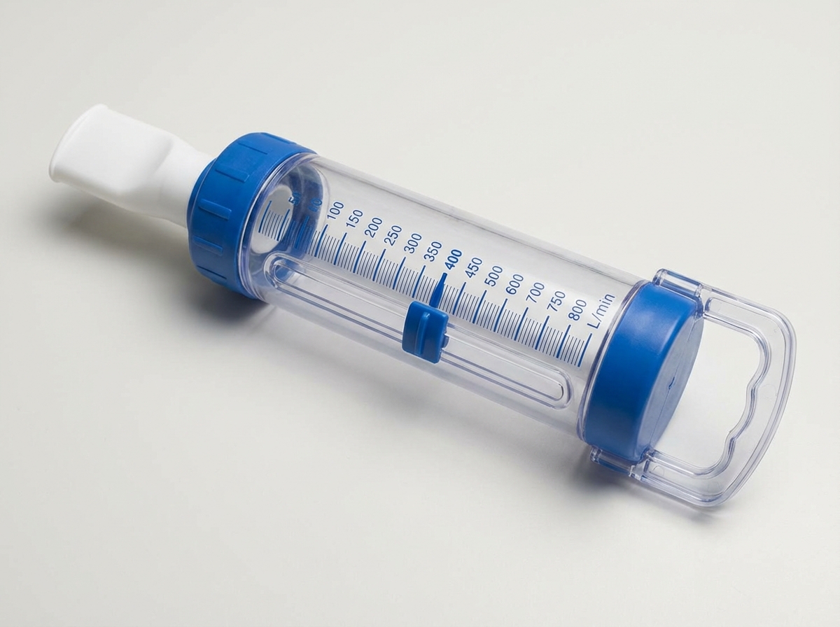
Identify the instrument: (Recent Neet Pattern 2016-17)

What is the diagnosis if a patient can only see 3 green dots on the Worth 4 Dot test?
Explanation: ***Snellen's chart*** - The image displays a series of rows with **optotypes (letters)** that decrease in size, which is characteristic of a **Snellen chart**. - This chart is specifically used to measure **visual acuity** by determining the smallest row of letters a person can read at a specific distance. *Ishihara's chart* - **Ishihara charts** are used to test for **color blindness** and consist of plates with colored dots that form numbers or patterns, which are not depicted in the image. - The chart shown focuses on letter recognition and size differentiation, not color perception. *Farnsworth-Munsell hue test* - The **Farnsworth-Munsell hue test** is used for precise **color discrimination** and involves arranging caps of varying hues in a continuum. - This test is distinct from the letter-based visual acuity chart shown in the image. *Pelli-Robson chart* - The **Pelli-Robson chart** is used to measure **contrast sensitivity**, presenting letters of a constant size but decreasing contrast. - While it uses letters, the chart in the image has letters of decreasing size, indicating a visual acuity test rather than contrast sensitivity.
Explanation: ***Correct Option: 5 minutes*** - The **entire optotype (letter)** of a Snellen's chart subtends an angle of **5 minutes of arc** at the nodal point of the eye when viewed from the standard testing distance - This is the fundamental design principle of the Snellen chart for visual acuity testing - At 6 meters (20 feet), a letter on the 6/6 (20/20) line subtends 5 minutes of arc in total height and width - This standardization allows for consistent visual acuity measurement across different testing scenarios *Incorrect: 1 minute* - This is the angle subtended by **each component part** of the letter (e.g., the width of a stroke or gap between strokes) - Not the angle for the entire letter - Used to define the detail that must be resolved for normal vision *Incorrect: 3 minutes* - No standard optical principle supports this value - Not related to Snellen chart design parameters *Incorrect: 10 minutes* - This angle is too large for standard Snellen chart optotypes - Would indicate significantly reduced visual acuity requirements - Not consistent with 6/6 (20/20) vision standards
Explanation: ***20 D*** - The image depicts a **binocular indirect ophthalmoscope (BIO)**, which is used for wide-field examination of the retina. - The **20 D lens** is the **most commonly used condensing lens** with a BIO for visualizing the entire retina. - It provides the **widest field of view** (approximately 45-50 degrees) with adequate magnification, making it ideal for comprehensive peripheral retinal examination. - Other standard BIO lenses include 14 D, 28 D, and 30 D, but **20 D offers the optimal balance** of field of view and magnification for complete retinal visualization. *58 D* - A **58 D lens** is not a standard condensing lens used with binocular indirect ophthalmoscopy. - While high-power lenses can be used with various ophthalmoscopic techniques, they are not conventional for BIO examination of the entire retina. *78 D* - A **78 D lens** is typically used with a **slit lamp biomicroscope** for a magnified view of the posterior pole and macular details. - It does not provide the wide-field view necessary for visualizing the **entire retina** when used with a BIO. - This lens is excellent for detailed examination of the optic disc and macula but has a limited field of view. *90 D* - A **90 D lens** is also primarily used with a **slit lamp biomicroscope** for excellent magnification of the macula and optic nerve head. - It provides a high-resolution, magnified view of a *limited area*, making it unsuitable for a comprehensive survey of the entire retina. - Like the 78 D, it's designed for detailed central retinal examination, not peripheral screening.
Explanation: ***Astigmatism*** - The image shows that light rays from a single point source are **not focused at a single point** on the retina, but rather spread across multiple focal points or a line. This pattern is characteristic of **astigmatism**, where the eye's cornea or lens has an irregular curvature. - Specifically, rays 1 and 2 converge at one point, while rays 3 and 4 converge at a different point, indicating that the eye has different refractive powers along different meridians, which defines **astigmatism**. *Myopia* - In **myopia (nearsightedness)**, light rays from a distant object focus **in front of the retina**, not at multiple planes or lines as depicted. - Myopia would show all rays converging to a single point before reaching the retina, resulting in a blurry image. *Hypermetropia* - In **hypermetropia (farsightedness)**, light rays from a distant object focus **behind the retina**, assuming the eye is unaccommodated. - This condition would show the rays converging to a single point beyond the retinal plane, not divergent focal points within the eye. *Presbyopia* - **Presbyopia** is an age-related condition where the eye's **lens loses its flexibility**, making it difficult to focus on nearby objects. - It primarily affects near vision and does not typically involve the multiple focal points for distant vision as shown in the image.
Explanation: ***A = Lister perimeter, B = Goldmann perimeter*** - Image A depicts a **Lister perimeter**, a type of **manual kinetic perimeter** used for visual field testing, characterized by its curved arm and a system for target presentation. - Image B shows a **Goldmann perimeter**, which is also a **manual kinetic perimeter** but features a large, bowl-shaped screen for testing, allowing for a broader and more controlled visual field assessment. *A = Goldmann perimeter, B = Lister perimeter* - This option incorrectly identifies image A as a **Goldmann perimeter** and image B as a **Lister perimeter**. - The distinct physical characteristics of each instrument, such as the curved arm of the Lister perimeter versus the bowl of the Goldmann perimeter, differentiate them. *A = Lister perimeter, B = Bjerrum's screen* - While image A is correctly identified as a **Lister perimeter**, image B is incorrectly identified as a **Bjerrum's screen**. - A **Bjerrum's screen** is a simpler, flat tangent screen used for central visual field testing, not the bowl-shaped instrument shown in image B. *A = Goldmann perimeter, B = Bjerrum screen* - This option incorrectly identifies image A as a **Goldmann perimeter** and image B as a **Bjerrum screen**. - The appearances of both the **Goldmann perimeter** and the **Bjerrum screen** do not match the instruments in images A and B, respectively.
Explanation: ***Maddox rod*** - The image displays a **Maddox rod**, characterized by a series of parallel, high-plus cylinders (rods) typically embedded in a red plastic disk with a handle. - This instrument is used to dissociate the eyes and convert a point source of light into a **line of light**, which is crucial for detecting and measuring heterophorias (latent deviations) and heterotropias (manifest deviations). *Maddox wing* - The **Maddox wing** is a different device used for measuring horizontal and vertical phorias at near. It consists of a septal plate that separates the visual fields of the two eyes, allowing the patient to see a set of scales with one eye and an arrow with the other. - It does not have the characteristic red parallel rods seen in the image. *Red green glasses* - **Red-green glasses** (or red-green anaglyph glasses) are used in various vision tests, such as stereopsis testing or some forms of vision therapy. - They selectively filter light, allowing one eye to see through a red filter and the other through a green filter, which is distinct from the multiple parallel rods shown. *Bagolini's striated glasses* - **Bagolini's striated glasses** are nearly plano lenses with very fine striations, designed to cause minimal disruption to vision while creating a streak of light from a point source. - They are primarily used to assess the presence and type of anomalous retinal correspondence and gross diplopia, and they do not have the prominent red rods as depicted.
Explanation: ***Bifocal glasses for presbyopia*** - The image clearly shows spectacle lenses with a visible **horizontal line separating two distinct optical powers**, which is characteristic of **bifocal lenses**. - **Presbyopia** is the condition where the eye's lens loses its ability to focus on near objects, requiring a different optical correction for near vision separate from distance vision, precisely what bifocals provide. *Progressive glasses for presbyopia* - **Progressive lenses** offer a gradual change in optical power from distance to near vision without a visible dividing line, unlike the spectacles shown. - They provide a continuous range of focus, but the absence of a visible segment in progressive lenses differentiates them from bifocals. *Bifocals for paediatric pseudo-phakia* - While pediatric pseudophakia (presence of an intraocular lens in a child) might require bifocals, the question asks for the primary use of the *pictured* bifocals, which commonly address **age-related presbyopia**. - Additionally, pseudophakia itself doesn't inherently imply a need for bifocals unless there's an accommodative issue, which is more typically associated with adult presbyopia. *Bifocals for adult aphakia* - **Aphakia** is the absence of the natural lens in the eye, which requires strong corrective lenses. While bifocals can be used in aphakia to provide both distance and near correction, the pictured bifocals are a standard design most commonly associated with correcting **presbyopia** in the general population. - Aphakic corrections generally involve much higher power lenses, which might appear thicker or have different characteristics than the standard bifocal shown.
Explanation: ***Mini-Wright peak flow meter*** - The image displays a **Mini-Wright peak flow meter**, identifiable by its characteristic tube for blowing into, connected to a plastic casing with a scale - This device is used to measure **Peak Expiratory Flow Rate (PEFR)**, which is crucial for monitoring conditions like **asthma** - The distinctive design with a mouthpiece, moving indicator, and numbered scale is pathognomonic of this device *Spirometer* - A spirometer is a larger device that measures multiple lung volumes and capacities (FVC, FEV1, etc.) - It typically has more complex components including a computerized display or graph paper recorder - Used for comprehensive pulmonary function testing, not just peak flow measurement *Incentive spirometer* - An incentive spirometer is used for post-operative breathing exercises - It has a different design with balls or pistons that rise with inhalation to provide visual feedback - Used for lung expansion therapy, not for measuring PEFR *Wright nebulizer* - A Wright nebulizer is used for administering aerosolized medications - It has a reservoir for liquid medication and produces a fine mist - Not a measurement device but a therapeutic delivery system
Explanation: ***Glover's Bulldog Clamp*** - The image displays a **surgical clamp** with characteristic **U-shaped jaws** that are relatively short and atraumatic - This specific design is consistent with a **Glover's bulldog clamp**, which is used to temporarily occlude blood vessels during vascular surgical procedures - The **spring-loaded mechanism** and **blunt, cushioned jaws** prevent vessel damage during temporary occlusion *Kocher's Clamp* - This is a **hemostatic forceps** with transverse serrations and interlocking teeth at the tip - Unlike the bulldog clamp, Kocher's clamp has **finger rings** and is used for grasping and holding tissue firmly *Allis Tissue Forceps* - Features **multiple interlocking teeth** arranged in rows for grasping delicate tissue - Has **ratcheted finger rings** for maintaining grip, unlike the spring mechanism shown *Babcock Forceps* - Has **fenestrated, rounded, non-traumatic jaws** designed for grasping hollow organs - Characterized by **wider, more rounded tips** than the narrow U-shaped jaws in the image
Explanation: ***Right eye suppression*** - Seeing **three green dots** exclusively indicates that the patient is only perceiving input from the **left eye**. - In the Worth 4 Dot test, the **left eye** (viewing through a green filter) sees **three green dots**: the white dot at the top (which appears green through the filter) plus the two lateral green dots. - The **right eye** (viewing through a red filter) normally sees **two red dots**: the white dot at the top (which appears red) plus the red dot at the bottom. - Since the patient sees only **three green dots**, the visual input from the **right eye is being suppressed**. *Crossed diplopia* - **Crossed diplopia** (heteronymous diplopia) occurs when the image from the right eye is perceived to the left of the image from the left eye. - This typically occurs with **exotropia** (divergent strabismus) and would result in seeing **five or more dots** (patient perceives both eyes' images but misaligned), not just three green. *Uncrossed diplopia* - **Uncrossed diplopia** (homonymous diplopia) occurs when the image from the right eye is perceived to the right of the image from the left eye. - This is usually associated with **esotropia** (convergent strabismus) and would also lead to the perception of **five or more dots** (both eyes' images perceived but misaligned), not only three green dots. *Left eye suppression* - If there were **left eye suppression**, the patient would see **two red dots** from the right eye only (the white dot appearing red plus the red dot at the bottom). - Seeing **three green dots** confirms the **left eye input is dominant** and the **right eye is suppressed**.
Physical Optics
Practice Questions
Geometric Optics
Practice Questions
Optical System of Eye
Practice Questions
Visual Acuity and Contrast Sensitivity
Practice Questions
Refractive Errors
Practice Questions
Accommodation and Presbyopia
Practice Questions
Optical Instruments
Practice Questions
Lenses and Prisms
Practice Questions
Retinoscopy
Practice Questions
Subjective Refraction
Practice Questions
Contact Lens Optics
Practice Questions
Wavefront Technology
Practice Questions
Get full access to all questions, explanations, and performance tracking.
Start For Free