
Optics and Refraction — MCQs

On this page
What is the approximate area of the fundus that can be visualized with a direct ophthalmoscope?
Aniseikonia means:
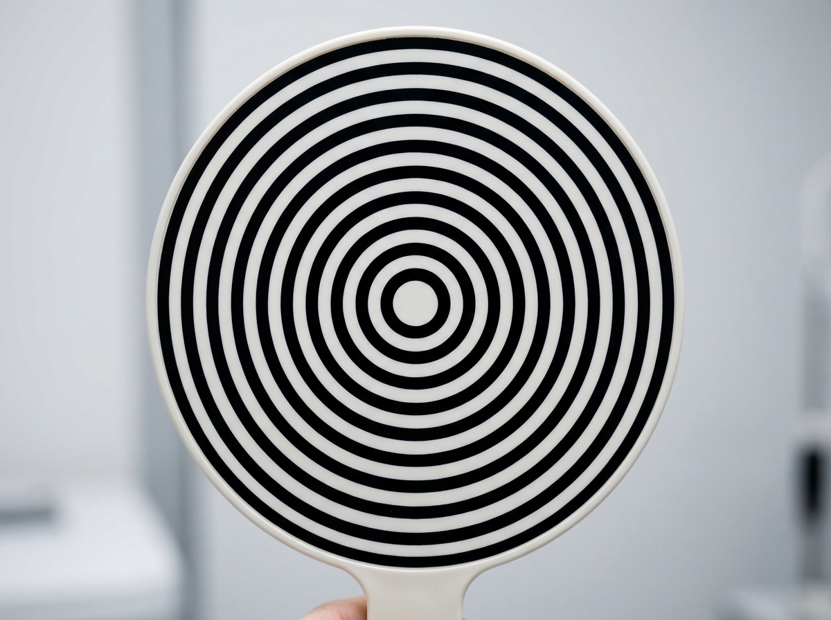
The instrument shown below is:
A 35-year-old hypermetrope is using 1.50 D sphere in both eyes. When his glasses slip downward on his nose, how does his near vision change?
What is the treatment for anisometropia?
All of the following are true about direct ophthalmoscopy except:
At what distance is direct ophthalmoscopy typically performed?
What is the distance between the nodal point and the cornea in Listing's Reduced eye?
What is the axial length of the reduced eye from the anterior surface of the cornea to the retina?
A person comes for a routine eye check-up. On Snellen's chart, they can read 6/6. At what distance should this person be able to read 6/24?
Practice by Chapter
Physical Optics
Practice Questions
Geometric Optics
Practice Questions
Optical System of Eye
Practice Questions
Visual Acuity and Contrast Sensitivity
Practice Questions
Refractive Errors
Practice Questions
Accommodation and Presbyopia
Practice Questions
Optical Instruments
Practice Questions
Lenses and Prisms
Practice Questions
Retinoscopy
Practice Questions
Subjective Refraction
Practice Questions
Contact Lens Optics
Practice Questions
Wavefront Technology
Practice Questions
Want unlimited practice?
Get full access to all questions, explanations, and performance tracking.
Scan to download app