
Optics and Refraction — MCQs

On this page
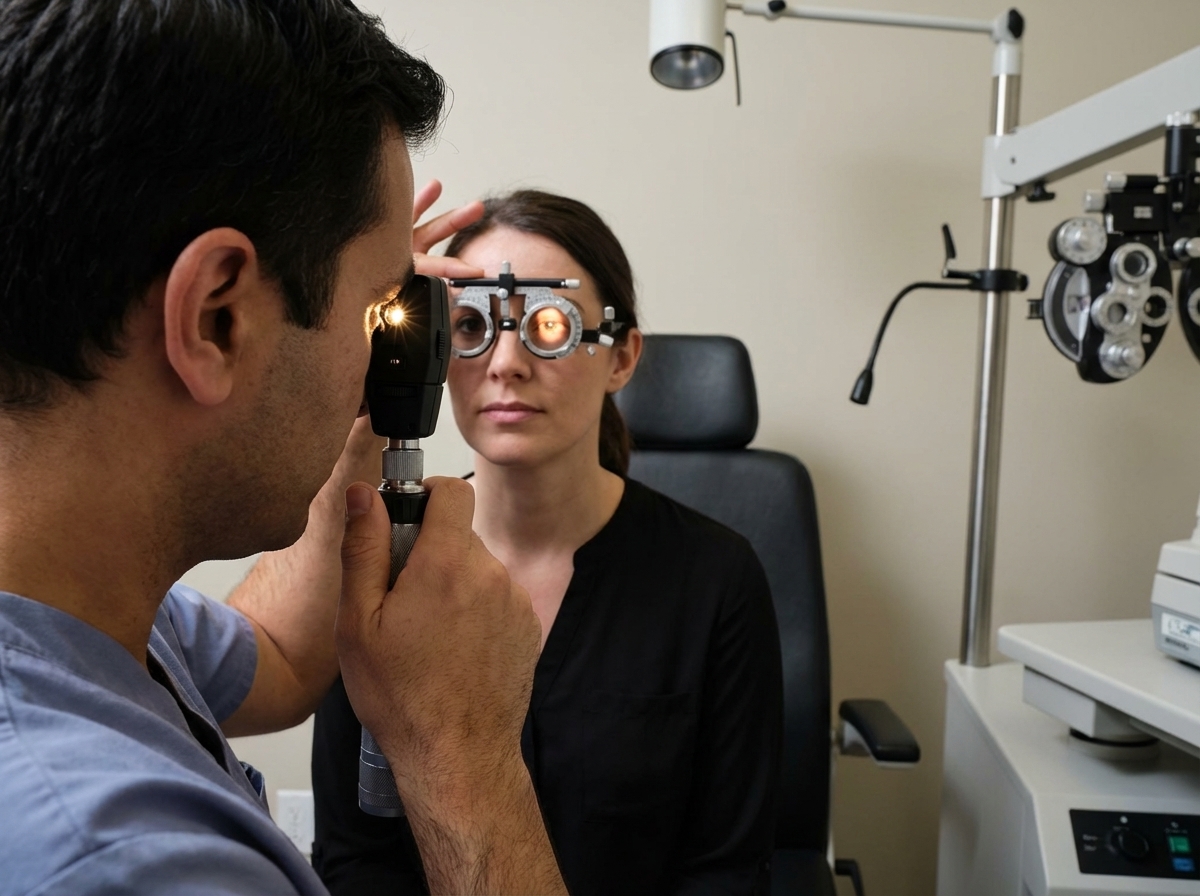
Name the procedure shown in the image?
The near point of the eye varies with which of the following factors?
What is the normal power of a reduced eye in diopters?
Which one of the following is a serious complication of degenerative myopia?
What is the approximate refractive power of the cornea?
Presbyopia occurs as a result of which of the following changes?
All of the following conditions can be diagnosed on distant direct ophthalmoscopy except?
Snellen's chart is based on which visual function?
Each layer of the lens has a role in the refractive index of the entire lens. Which component has the maximum refractive index?
Which mydriatic is used for refraction in infants?
Practice by Chapter
Physical Optics
Practice Questions
Geometric Optics
Practice Questions
Optical System of Eye
Practice Questions
Visual Acuity and Contrast Sensitivity
Practice Questions
Refractive Errors
Practice Questions
Accommodation and Presbyopia
Practice Questions
Optical Instruments
Practice Questions
Lenses and Prisms
Practice Questions
Retinoscopy
Practice Questions
Subjective Refraction
Practice Questions
Contact Lens Optics
Practice Questions
Wavefront Technology
Practice Questions
Want unlimited practice?
Get full access to all questions, explanations, and performance tracking.
Scan to download app