Argon laser trabeculoplasty is done in:
Uncontrolled hypertension may cause which of the following complications in cataract surgery:
Laser iridotomy is done in?
A crucial step in the phacoemulsification procedure for a 70-year-old man scheduled for cataract surgery is which of the following?
A 56-year-old patient presents after 3 days of cataract surgery with a history of increasing pain and diminished vision after an initial improvement. The most likely cause would be –
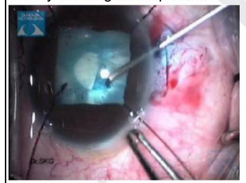
Identify the surgical step shown in the image given below

Expulsive hemorrhage in cataract surgery is from?
In which of the following conditions does IOL implantation after cataract surgery require the greatest caution and specialized management?
What is the preferred surgical technique for traumatic cataract in children?
Which of the following procedures is not typically covered by the National Programme for Control of Blindness (NPCB) for reimbursement of surgery done by a non-governmental organization (NGO) eye hospital?
Explanation: ***Open angle glaucoma*** - **Argon laser trabeculoplasty (ALT)** is primarily used in **open-angle glaucoma** to improve aqueous humor outflow through the **trabecular meshwork**. - It creates **small burns** in the trabecular meshwork, increasing permeability and lowering **intraocular pressure (IOP)**. *Secondary glaucoma* - This is a broad category of glaucoma caused by other eye conditions or systemic diseases, and the specific treatment depends on the underlying etiology. - While ALT might be considered in some types of **secondary open-angle glaucoma**, it is not a primary or universal treatment for all secondary glaucomas. *Angle recession glaucoma* - This type of glaucoma occurs after blunt trauma to the eye, causing a tear in the **ciliary body** and widening of the **ciliary body band**. - ALT is generally **ineffective** in angle recession glaucoma because the damage to the trabecular meshwork is structural and not amenable to laser modification. *Angle closure glaucoma* - In **angle-closure glaucoma**, the iris blocks the drainage angle, preventing aqueous outflow. - Treatment typically involves **laser peripheral iridotomy** to create a hole in the iris, or surgical procedures, to open the angle, rather than laser trabeculoplasty.
Explanation: ***Suprachoroidal hemorrhage*** - **Uncontrolled hypertension** significantly increases the risk of **suprachoroidal hemorrhage** during cataract surgery due to fragile blood vessels and elevated intraoperative blood pressure. - This complication can lead to acute, severe pain, vision loss, and globe rupture, often requiring immediate surgical intervention. *Retinal detachment* - While a serious complication of ocular surgery, **retinal detachment** is not directly caused by uncontrolled hypertension during cataract surgery. - It is more commonly associated with posterior capsular rupture, vitreous loss, or high myopia. *Glaucoma* - **Glaucoma** is a chronic condition characterized by optic nerve damage, often due to elevated intraocular pressure, and is not an acute complication of uncontrolled hypertension during cataract surgery. - While hypertension is a risk factor for certain types of glaucoma, it does not directly cause an acute glaucomatous event during the procedure. *Endophthalmitis* - **Endophthalmitis** is a severe infection of the intraocular fluids and tissues, typically occurring post-operatively. - It is primarily caused by bacterial or fungal contamination during or after surgery and is not directly linked to uncontrolled hypertension.
Explanation: ***Angle closure glaucoma*** - **Laser iridotomy** creates a small hole in the iris, allowing aqueous humor to flow directly from the posterior to the anterior chamber, thus relieving pupillary block and opening the angle. - This procedure is the definitive treatment to prevent further **angle closure attacks** and is also used prophylactically in eyes at risk. *Open angle glaucoma* - This condition involves an **open angle** but impaired outflow of aqueous humor through the **trabecular meshwork**. - Laser iridotomy is not indicated as it does not address the primary outflow obstruction in the trabecular meshwork. *Pigmentary glaucoma* - This is a type of **open-angle glaucoma** caused by pigment dispersion that clogs the trabecular meshwork, leading to increased intraocular pressure. - While pigment can be released from the iris, the primary issue is the **trabecular meshwork obstruction**, which is not directly resolved by iridotomy. *None of the options* - This option is incorrect because **angle closure glaucoma** is a clear indication for laser iridotomy.
Explanation: ***Continuous curvilinear capsulorrhexis*** - This step creates a smooth, continuous, and appropriately sized opening in the **anterior lens capsule**, which is crucial for the safe and effective removal of the cataractous lens material. - A well-executed capsulorrhexis ensures the **intraocular lens (IOL)** can be stably implanted within the capsular bag, minimizing complications like IOL decentering. *Scleral buckling* - **Scleral buckling** is a surgical procedure primarily used to repair **retinal detachments**, not for cataract removal. - It involves placing a silicone band on the outer surface of the sclera to indent the eye wall, supporting the retina. *Corneal transplantation* - **Corneal transplantation** (keratoplasty) is performed to replace a diseased or damaged cornea, typically for conditions like **keratoconus** or corneal scarring. - It is not a component of routine cataract surgery. *Trabeculectomy* - **Trabeculectomy** is a surgical procedure to treat **glaucoma** by creating a new drainage pathway for aqueous humor, thereby reducing intraocular pressure. - It is unrelated to the process of cataract extraction.
Explanation: ***Endophthalmitis*** - The presentation of **increasing pain** and **diminished vision** within days of cataract surgery, following an initial improvement, is highly suggestive of acute **postoperative endophthalmitis**. - This severe **intraocular inflammation** is often caused by bacterial infection introduced during surgery, leading to rapid vision loss if not treated promptly. *Posterior capsular opacification* - This condition typically presents weeks to months or even years after cataract surgery, not within 3 days. - It usually causes **gradual blurring of vision** without pain, unlike the acute symptoms described. *Central retinal vein occlusion* - This condition presents with **sudden, painless vision loss** and a characteristic appearance on fundoscopy (e.g., "blood and thunder" retina). - It is not directly related to cataract surgery and would not typically cause increasing pain. *Retinal detachment* - Symptoms usually include **new floaters**, **flashes of light**, and a **"curtain" or "shadow"** over the field of vision, often developing suddenly and progressing. - While it causes vision loss, it is typically painless and not a direct complication presenting with pain within 3 days post-surgery, especially after initial improvement.
Explanation: ***Lens aspiration*** - The image shows a **phacoemulsification handpiece** (the instrument with the shining tip and central bore tube) actively fragmenting and aspirating the lens material, indicated by the cloudy material being removed. - This step is part of cataract surgery where the cataractous lens material is removed from the eye. *Capsulorrhexis* - This involves creating a **continuous curvilinear tear** in the anterior lens capsule, typically done at the beginning of cataract surgery. - The image does not show a tearing or incising action on the capsule; instead, it depicts material removal. *Hydrodissection* - This step involves injecting a **fluid wave** between the lens capsule and the lens cortex to separate them, facilitating nuclear rotation and removal. - The image depicts the removal of lens material, not the injection of fluid to separate layers. *Intraocular lens implantation* - This step involves inserting the **artificial lens** into the capsular bag after the cataractous lens has been removed. - The visual cues in the image indicate material removal and emulsification, not the insertion of a new lens.
Explanation: ***Ciliary artery*** - Expulsive hemorrhage is a rare but devastating complication, typically resulting from the rupture of a **posterior ciliary artery** within the choroid. - This arterial rupture leads to a sudden, massive increase in intraocular pressure and extrusion of intraocular contents. *Vortex vein* - **Vortex veins** drain the choroid, and while their rupture could lead to hemorrhage, it is less likely to cause the highly pressurized, expulsive nature of a choroidal hemorrhage. - Hemorrhage from a vortex vein is generally less severe and less rapid in onset compared to arterial bleeding. *Choroidal vein* - **Choroidal veins** are part of the venous drainage system; bleeding from these vessels would typically be lower pressure and less likely to cause an expulsive hemorrhage. - Venous bleeds are generally slower and do not generate the rapid, violent pressure increase characteristic of expulsive hemorrhage. *None of the options* - This option is incorrect because the rupture of a ciliary artery is the direct cause of expulsive hemorrhage. - The other options are incorrect for the reasons stated above.
Explanation: ***Juvenile rheumatoid arthritis*** - Patients with **juvenile rheumatoid arthritis (JRA)**, particularly those with **pauciarticular JRA** and **ANA positivity**, are at high risk for developing chronic uveitis, which can lead to significant cataract formation and severe postoperative complications. - Due to the high risk of severe postoperative inflammation, glaucoma, and vision loss, IOL implantation in JRA patients requires extensive preoperative optimization of inflammation and careful intraoperative/postoperative management. *Fuchs' heterochromic iridocyclitis* - This condition presents with chronic, low-grade, **non-granulomatous anterior uveitis** and often leads to cataract formation. - While IOL implantation in these patients is generally well-tolerated, it does not pose the same high risk of severe postoperative inflammation and complications as seen in JRA-associated uveitis. *Psoriatic arthritis* - Psoriatic arthritis can be associated with acute anterior uveitis, but it typically presents as an acute, intermittent inflammation. - The risk of chronic, severe uveitis leading to complex cataract surgery and significant postoperative complications is not as consistently high or as severe as in JRA. *Reiter's syndrome* - Reiter's syndrome (now part of **reactive arthritis**) is another seronegative spondyloarthropathy that can cause acute anterior uveitis. - Similar to psoriatic arthritis, the uveitis is usually acute and self-limiting, and while ocular inflammation needs to be controlled, the risk profile for IOL implantation is not as challenging as in JRA.
Explanation: ***Lensectomy (Lensectomy-Vitrectomy)*** - **Lensectomy with anterior vitrectomy** is the preferred surgical approach for traumatic cataracts in children - Traumatic cataracts often have **compromised capsular integrity** and zonular weakness, making ECCE unsuitable - The procedure removes lens material and posterior capsule, significantly reducing the **high risk of posterior capsule opacification (PCO)** in children - **Primary IOL implantation** may be performed if the child is >2 years old and capsular support is adequate; otherwise, aphakic correction with contact lenses or secondary IOL is planned - Modern technique provides better long-term visual outcomes in pediatric trauma cases *Extracapsular Cataract Extraction (ECCE) + IOL* - **ECCE is largely outdated** for pediatric traumatic cataracts due to several limitations - Traumatic cataracts frequently have **capsular damage and zonular dehiscence**, making ECCE technically difficult and risky - Children have very high rates of **posterior capsule opacification (PCO)** with retained posterior capsule, requiring multiple YAG procedures - Cannot adequately address **vitreous complications** often associated with traumatic cataracts *Contact lenses for vision correction post-surgery* - Contact lenses are a **rehabilitation option**, not a surgical technique - Used for aphakic correction when primary IOL is not implanted (very young children or inadequate capsular support) - This addresses optical correction after surgery, not the surgical approach itself *Glasses for vision correction post-surgery* - Glasses are a **rehabilitation option**, not a surgical technique - Can provide aphakic correction but result in significant magnification and poor cosmesis - Less preferred than contact lenses for aphakic children due to optical limitations
Explanation: ***Syringing and probing of the nasolacrimal duct*** - While important for lacrimal drainage issues, procedures like **syringing and probing** are generally considered minor and less vision-restoring compared to the major surgeries targeted by the **NPCB**. - The **NPCB** focuses on interventions for leading causes of blindness, primarily **cataract** and other significant vision-threatening conditions, which this procedure typically isn't. *Cataract surgery* - **Cataract surgery** is a cornerstone of the **NPCB's** efforts, as cataracts are the leading cause of reversible blindness. - Reimbursement for **cataract surgery** is a primary objective to improve access and reduce the burden of blindness. *Pan retinal photocoagulation for diabetic retinopathy* - **Diabetic retinopathy** is a major cause of preventable blindness, and **pan retinal photocoagulation (PRP)** is a key intervention to preserve vision. - The **NPCB** includes procedures for **diabetic retinopathy** management due to its significant public health impact. *Trabeculectomy surgery* - **Trabeculectomy** is a surgical procedure for **glaucoma**, which is another significant cause of irreversible blindness. - The **NPCB** includes interventions for **glaucoma** given its severe vision-threatening nature and the need for surgical management in many cases.
Get full access to all questions, explanations, and performance tracking.
Start For Free