Premedication is prescribed to – a) Allay anxiety b) Make the patient asleep before coming for operation c) Reduce the dose of induction agents d) Produce amnesia
Which pre-operative investigation is recommended before surgical procedures in a patient on warfarin therapy?
Retrobulbar injection of xylocaine blocks all of the following except:
A patient aged 28 years, was given epidural anesthesia with 15 ml of 1.5% Lignocaine with adrenaline for hernia surgery. He developed hypotension, respiratory arrest and became unconscious within 3 minutes, most probable cause will be:-
Local anaesthetic usually used for retrobulbar block -
What is the maximum dose of plain lignocaine (in mg) for adults?
A 5 year old healthy child is undergoing strabismus surgery with a Laryngeal mask airway in place. Thirty minutes into the procedure, his heart rate is 60 bpm, blood pressure is 90/60 mmHg, and oximeter shows 98% saturation. The next step in management is
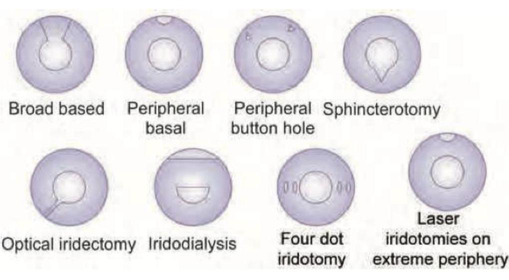
The type of iridectomy shown in the image is:

Which one of the following is a branch of the first part of the maxillary artery?
Identify the ophthalmic instrument used for measuring heterophoria and heterotropia.
Explanation: ***Allay anxiety*** - Premedication frequently includes anxiolytic agents like **benzodiazepines** to calm the patient before surgery. - Reducing anxiety helps in achieving a smoother induction of anesthesia and can improve the patient's overall experience. *Reduce the dose of induction agents* - While some premedication agents like **opioids** or sedatives can have an anesthetic-sparing effect, this is a secondary benefit, not the primary goal. - The main aim is patient comfort and psychological preparation, not primarily dose reduction. *Produce amnesia* - Amnesia, particularly **anterograde amnesia**, is a desirable side effect of some premedication drugs like **midazolam**. - However, it's a consequence of the anxiolytic effect rather than the sole or primary reason for prescribing premedication. *Make the patient asleep before coming for operation* - While some premedication agents can cause **somnolence** or light sleep, the goal is not to have the patient fully asleep before entering the operating room. - The primary aim is to make the patient relaxed and comfortable, not unconscious.
Explanation: ***International Normalized Ratio (INR)*** - The **INR** is specifically used to monitor the effectiveness of **warfarin** therapy, as it standardizes the prothrombin time (PT) for variations in thromboplastin reagents. - Before surgery, an INR measurement helps assess the patient's **coagulation status** and guides decisions on temporary cessation or bridging therapy to minimize bleeding risk. *Partial Thromboplastin Time (PTT)* - **PTT** primarily measures the **intrinsic and common pathways** of coagulation and is used to monitor **heparin** therapy, not warfarin. - While prolonged in some bleeding disorders, it is not the standard test for assessing warfarin's anticoagulant effect. *Clotting Time* - **Clotting time** is a very general and less precise measure of overall coagulation that is **rarely used** in modern clinical practice due to its low sensitivity and specificity. - It does not offer sufficient detail or standardization to guide pre-operative management for patients on warfarin. *Differential Count* - A **differential count** measures the different types of **white blood cells** within a blood sample and is used to diagnose infections, inflammatory conditions, or hematologic disorders. - It provides no information about a patient's coagulation status or the effects of anticoagulant medications like warfarin.
Explanation: ***4th cranial nerve*** - The **trochlear nerve (CN IV)** innervates the **superior oblique muscle** and has a unique superior and anterior course in the orbit, making it less accessible to retrobulbar injections. - Its location relative to the muscle cone and globe means a retrobulbar injection, which typically aims to block nerves within the muscle cone, often misses it. *3rd cranial nerve* - The **oculomotor nerve (CN III)** supplies most of the extraocular muscles and travels within the **muscle cone**, where retrobulbar anesthetic is deposited. - It is reliably blocked by a retrobulbar injection, causing akinesia of the muscles it innervates. *6th cranial nerve* - The **abducens nerve (CN VI)** innervates the **lateral rectus muscle** and is located within the **muscle cone**, making it susceptible to retrobulbar block. - Anesthetic diffusion within the cone effectively blocks this nerve, leading to paralysis of the lateral rectus. *Ciliary nerves and ciliary ganglion* - The **short ciliary nerves** and **ciliary ganglion** are located in the retrobulbar space, typically within the muscle cone or close to it. - Anesthetic solution injected retrobulbarly readily diffuses to these structures, blocking sensory input from the cornea and iris, as well as parasympathetic innervation to the pupil and ciliary body.
Explanation: ***Intravascular injection of Lignocaine*** - Rapid onset (within 3 minutes) of **hypotension**, **respiratory arrest**, and **unconsciousness** after an epidural injection strongly indicates systemic toxicity from intravascular local anesthetic injection. - The large volume (15 mL) and concentration (1.5%) of lignocaine, especially with adrenaline, when injected directly into the bloodstream, can quickly lead to **central nervous system (CNS) depression** and cardiovascular collapse. *High spinal block* - A **high spinal block** typically results from a local anesthetic spreading too high in the intrathecal space, leading to widespread sympathetic blockade and paralysis of respiratory muscles. - While it causes hypotension and respiratory depression, the rapid onset and immediate unconsciousness, without prior signs of extensive motor block ascending, make intravascular injection a more probable cause for such acute and severe symptoms. *Anaphylaxis to lignocaine* - Anaphylaxis to local anesthetics is rare and would typically present with **urticaria**, **angioedema**, **bronchospasm**, and widespread erythema, which are not described. - While anaphylaxis can cause hypotension and cardiovascular collapse, the rapid onset of CNS depression leading to unconsciousness is more characteristic of local anesthetic systemic toxicity. *Total spinal block* - A **total spinal block** occurs when a local anesthetic meant for the epidural space accidentally enters the subarachnoid space and diffuses extensively. - This results in profound **hypotension**, **bradycardia**, and **apnea** due to high sympathetic and somatic nerve blockade; however, unconsciousness typically ensues after significant hypotension and hypoperfusion, not as immediately and severely as seen with direct intravascular injection of a toxic dose.
Explanation: ***Bupivacaine*** - **Bupivacaine** is a long-acting local anaesthetic commonly chosen for retrobulbar blocks due to its prolonged duration of action, providing extended pain relief and akinesia. - Its slower onset compared to some other agents is often acceptable for ophthalmic procedures where prolonged block is more critical than rapid onset. *Prilocaine* - **Prilocaine** is an intermediate-acting local anaesthetic, generally having a shorter duration of action than bupivacaine, making it less ideal for procedures requiring sustained blockade. - High doses of prilocaine are associated with the risk of **methemoglobinemia**, which is a significant consideration in its use. *Tetracaine* - **Tetracaine** is primarily used as a topical anaesthetic, particularly for surface anaesthesia of the eye (e.g., prior to drops or contact lens insertion). - It is not typically used for injection in retrobulbar blocks due to its high systemic toxicity when administered via injection and its short duration of action. *Procaine* - **Procaine** is a short-acting ester-type local anaesthetic with a rapid onset but very brief duration of action. - Its short duration makes it unsuitable for retrobulbar blocks where prolonged akinesia and anaesthesia are desired for the surgical procedure.
Explanation: ***300 mg*** - The maximum recommended dose of **plain lidocaine** (without epinephrine) for adults is typically **300 mg** or 4.5 mg/kg, whichever is less. - Exceeding this dose can increase the risk of **systemic toxicity**, including central nervous system and cardiovascular effects. *500 mg* - This dose is generally considered the maximum for **lidocaine with epinephrine** in adults, as **epinephrine** causes vasoconstriction and delays systemic absorption of lidocaine. - For **plain lidocaine**, 500 mg would be an overdose and significantly increase the risk of toxicity. *700 mg* - This is well above the recommended maximum dose for both plain lidocaine and lidocaine with epinephrine, posing a **severe risk of toxicity**. - Such a high dose could lead to **seizures**, cardiac arrhythmias, and even **cardiac arrest**. *1000 mg* - This dose is extremely dangerous and far exceeds any recommended maximum for lidocaine, regardless of whether it contains epinephrine. - Administration of 1000 mg of lidocaine would almost certainly result in **severe and potentially fatal toxicity**.
Explanation: ***Inform surgeon, administer atropine*** - **Oculocardiac reflex** is a common complication during strabismus surgery, characterized by bradycardia, hypotension, and arrhythmias. The significant drop in heart rate (60 bpm in a 5-year-old child) and mild hypotension suggest this reflex. - The standard management for an oculocardiac reflex involves informing the surgeon to temporarily cease manipulation of the extraocular muscles and administering an **anticholinergic drug** like atropine to counteract the vagal stimulation. *Replace the LMA with endotracheal tube* - The patient's oxygen saturation of 98% indicates adequate ventilation and oxygenation with the LMA, so there is no immediate need for **airway intervention**. - Replacing the LMA with an endotracheal tube is a more invasive procedure and would not directly address the underlying cause of bradycardia, which is likely due to the oculocardiac reflex. *Nothing, this is normal for this child* - A heart rate of 60 bpm is **significantly low** for a 5-year-old child under anesthesia, as the expected heart rate for this age group is typically much higher (around 80-120 bpm). - Ignoring this bradycardia could lead to further compromise in **cardiac output** and tissue perfusion if not addressed promptly. *Increase FiO2 to 1.0* - The current oxygen saturation of 98% indicates **adequate oxygenation**, so increasing the FiO2 would not address the bradycardia or hypotension. - While maintaining good oxygenation is important, this step would not resolve the primary issue of an **oculocardiac reflex** causing vagal stimulation.
Explanation: ***Peripheral basal iridectomy*** - The image clearly depicts an iris with a small, circular opening located at its **periphery**, specifically at the base near the ciliary body. - This type of opening is characteristic of a **peripheral basal iridectomy**, which creates an alternative pathway for aqueous humor flow to relieve pupillary block, often associated with angle-closure glaucoma. *Sector iridectomy* - A **sector iridectomy** involves removing a full-thickness, wedge-shaped section of the iris that extends from the pupillary margin to the iris root, creating a keyhole-shaped pupil. - The image does not show a wedge-shaped defect extending to the pupil. *Button-hole iridectomy* - A **button-hole iridectomy** is typically a small, central opening in the iris that is completely surrounded by iris tissue, often performed for optical purposes. - The image shows a peripheral opening, not a central one. *Complete iridectomy* - A **complete iridectomy** implies the removal of the entire iris, or at least a very large portion, which would result in a highly enlarged and distorted pupil. - The image shows a small, localized opening, not extensive iris removal.
Explanation: ***Anterior tympanic*** - This artery arises from the **first part** (mandibular part) of the maxillary artery. - It supplies structures within the **tympanic cavity**, including the inner surface of the tympanic membrane. - Among the options listed, this is a **classic branch** consistently mentioned in anatomy texts. *Anterior ethmoidal* - This artery is a branch of the **ophthalmic artery**, which itself is a branch of the internal carotid artery, **not the maxillary artery**. - It supplies the **ethmoid air cells**, frontal sinus, and nasal cavity. - This is the **definitively incorrect option** as it does not arise from the maxillary artery at all. *Middle meningeal* - This artery **also arises from the first part** (mandibular part) of the maxillary artery. - It is a significant artery that supplies the **dura mater** and cranial bones. - While anatomically correct, **anterior tympanic** is the more specific answer being tested in this context. *Inferior alveolar* - This artery **also arises from the first part** (mandibular part) of the maxillary artery. - It descends to supply the **mandible**, its teeth, and the lower lip and chin. - While anatomically correct, it is not the best answer in this specific question context.
Explanation: ***Maddox rod*** - A **Maddox rod** consists of a series of parallel cylindrical lenses that converts a point source of light into a line, forcing the patient to dissociate the images seen by each eye. - This dissociation allows for the measurement of **heterophoria** (latent deviation) and **heterotropia** (manifest deviation or strabismus) by observing the position of the perceived line relative to a fixation light. *Maddox wing* - The Maddox wing is used to measure **heterophoria at near**, typically at 33 cm. - It presents separate images to each eye (a scale and arrows) and does not involve the principle of converting a point source to a line. *Maddox glass* - The term **Maddox glass** is not a standard ophthalmic instrument. - It might colloquially refer to a Maddox rod or a similar device, but it lacks the specific definition of the other options. *Red glasses* - **Red glasses** (or red filter) are used in various ophthalmic tests, often to create dissociation between the eyes or to test for suppression. - They do not, however, convert a point source of light into a line for the precise measurement of ocular deviation in the same way a Maddox rod does.
Get full access to all questions, explanations, and performance tracking.
Start For Free