
Oculoplasty — MCQs

On this page
Recurrent chalazion is predisposed to develop which of the following?
A 65-year-old woman presents with complaints of pain and swelling over the inner aspect of the right eye for the past two days. Examination reveals tenderness, edema, and redness over the medial canthus. Slight pressure over the area causes expression of purulent material. Visual acuity is normal. What is the most likely diagnosis?
A 60-year-old man presented with watering from his left eye for one year. Syringing revealed a patent drainage system. The rest of the ocular examination was normal. A provisional diagnosis of lacrimal pump failure was made. Confirmation of the diagnosis would be by:
The Schirmer's test is performed to evaluate the function of which gland?
Lipogranulomatous inflammation is characteristic of which of the following conditions?
A 60-year-old man presented with watering from his left eye for one year. Syringing revealed a patent drainage system. The rest of the ocular examination was normal. A provisional diagnosis of lacrimal pump failure was made. Confirmation of the diagnosis would be by:
What is the commonest tumor found at the inner canthus of the eye?
What is the most common cause of trichiasis?
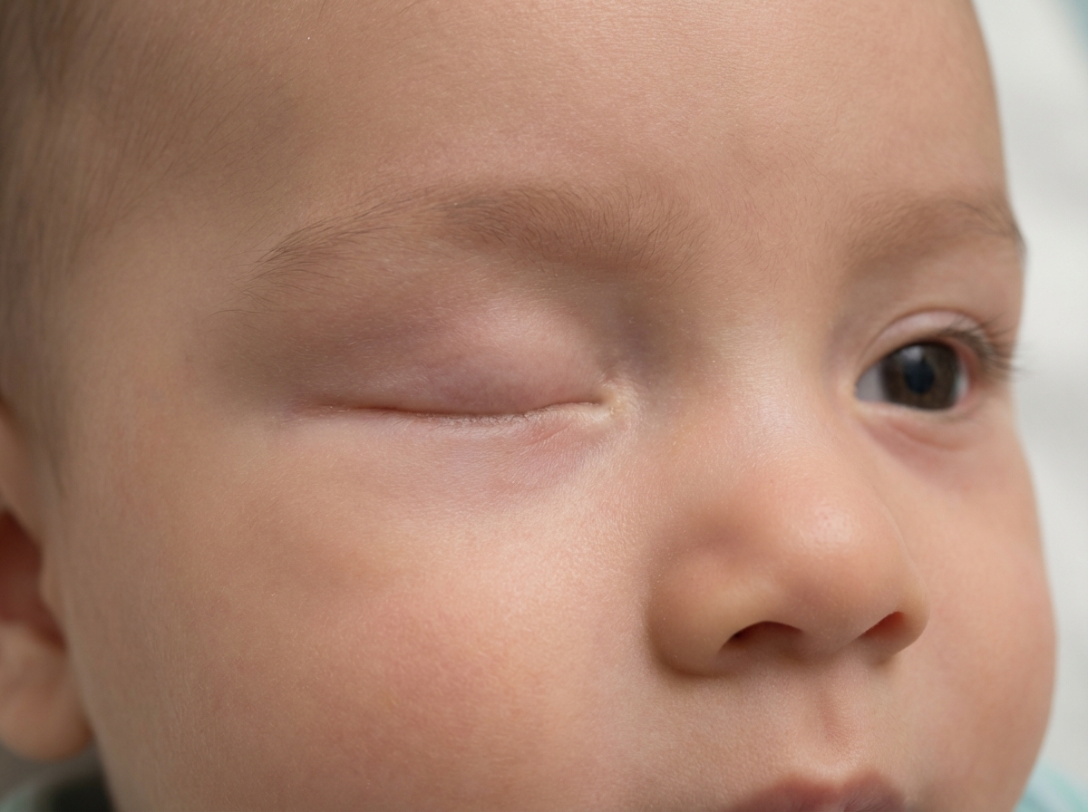
All of the following statements are true about the given condition except?
Probing and irrigation is not indicated in which of the following conditions?
Practice by Chapter
Eyelid Anatomy and Physiology
Practice Questions
Ptosis
Practice Questions
Entropion and Ectropion
Practice Questions
Eyelid Tumors
Practice Questions
Facial Nerve Palsy
Practice Questions
Blepharospasm and Hemifacial Spasm
Practice Questions
Blepharitis and Meibomian Gland Dysfunction
Practice Questions
Lacrimal System Disorders
Practice Questions
Orbital Inflammations
Practice Questions
Orbital Tumors
Practice Questions
Thyroid Eye Disease
Practice Questions
Anophthalmic Socket Management
Practice Questions
Want unlimited practice?
Get full access to all questions, explanations, and performance tracking.
Scan to download app