Facial nerve does not supply which structure of the face?
Unilateral frontal blisters with upper lid edema and conjunctivitis is seen in?
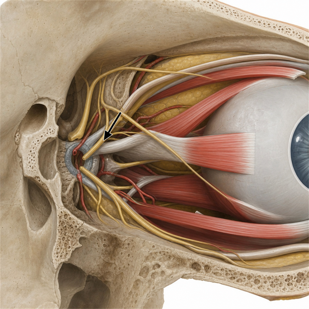
MC site of basal cell carcinoma of eyelid:
The infratentorial dura is supplied by branches of the ___?
Chronic granulomatous inflammation in upper lid (painless swelling) is characteristic of:
Muscle in the lid attached to posterior tarsal margin is:
A patient with ptosis has the upper 4 mm of cornea covered by the upper eyelid. What is the grade of ptosis?
Internal hordeolum is due to inflammation of-
Distichiasis is a condition characterized by:
The muscle first affected in thyroid ophthalmopathy is:
Explanation: ***Parotid gland*** - While the facial nerve (CN VII) passes *through* the parotid gland, it does not provide motor innervation to the gland itself. - The parotid gland receives parasympathetic innervation for **salivation** primarily from the **glossopharyngeal nerve (CN IX)** via the otic ganglion. *Posterior belly of digastric muscle* - The **facial nerve (CN VII)** provides motor innervation to the posterior belly of the digastric muscle. - This muscle is involved in **depressing the mandible** and **elevating the hyoid bone**. *Submandibular gland* - The facial nerve (CN VII) provides parasympathetic secretomotor innervation to the submandibular gland via the **chorda tympani** and submandibular ganglion. - This innervation controls **salivation** from the submandibular gland. *Auricular muscle* - The facial nerve (CN VII) supplies the **auricular muscles**, which are muscles of facial expression around the ear. - These muscles contribute to minor **ear movements**.
Explanation: ***Herpes Zoster Ophthalmicus*** - This condition is characterized by a **unilateral vesicular rash** (blisters) in the **trigeminal dermatome (V1)**, which includes the forehead and upper eyelid, along with significant **lid edema** and **conjunctivitis**. - **Hutchinson's sign** (lesions on the tip, side, or root of the nose) indicates a high risk of ocular involvement due to the nasociliary nerve innervation. *Acanthamoeba Keratitis* - This is an **amoebic infection** of the cornea typically associated with **contact lens wear** and often presents with severe pain and a **ring infiltrate** in the cornea. - It does not typically present with unilateral frontal blisters or significant lid edema. *Herpes Simplex* - Herpes simplex typically causes **recurrent corneal ulcers** (dendritic or geographic) and sometimes blepharitis, but not the widespread **unilateral frontal blisters** seen in the trigeminal distribution. - While it can cause conjunctivitis and lid edema, the pattern of skin lesions is the key differentiator. *Neuroparalytic Keratitis* - This condition results from **trigeminal nerve damage**, leading to corneal anesthesia and subsequent **trophic corneal ulceration**. - It presents primarily with **corneal findings** (epithelial defects, ulcers) due to impaired sensation and tear film stability, not initial vesicular skin lesions or prominent lid edema.
Explanation: ***Lower eyelid*** - The **lower eyelid** is the most common site for basal cell carcinoma (BCC) of the eyelid, accounting for approximately **50-60%** of all eyelid BCCs. - This high frequency is due to increased exposure to **UV radiation**, which is the primary risk factor for BCC development. - BCC often presents as a **pearly nodule** with telangiectasias and central ulceration, frequently found on the lower lid margin. *Medial canthus* - The medial canthus is the **second most common site**, accounting for approximately **25-30%** of eyelid BCCs. - Tumors in this area can be **more aggressive** and challenging to treat due to proximity to the lacrimal system and orbital structures. - Medial canthal BCCs may require more extensive surgical reconstruction. *Upper eyelid* - The upper eyelid accounts for only **10-15%** of eyelid BCCs, making it significantly **less common** than the lower eyelid. - This is due to **less direct sun exposure** compared to the lower lid, as the upper lid is often shaded by the brow. *Outer canthus* - The outer (lateral) canthus is the **least common site**, accounting for only about **5%** of eyelid BCCs. - Tumors here may present with similar features but are much less frequently encountered than those on the lower lid or medial canthus.
Explanation: ***Upper cervical spinal nerves and vagus nerve*** - The **infratentorial dura mater**, particularly the posterior fossa, receives its sensory innervation primarily from the **recurrent meningeal branches** of the upper cervical spinal nerves (C1-C3), which ascend through the foramen magnum. - The **vagus nerve (CN X)** also contributes to the sensory supply of the infratentorial dura, specifically to the posterior fossa, through its sensory branches. *Accessory nerve and upper cervical nerves* - The **accessory nerve (CN XI)** is primarily a motor nerve, responsible for innervating the sternocleidomastoid and trapezius muscles, and does not directly supply the dura mater. - While upper cervical nerves do contribute, the **vagus nerve** is also a significant contributor to infratentorial dural innervation. *Only vagus nerve* - While the **vagus nerve (CN X)** does contribute to the sensory innervation of the infratentorial dura, it is not the sole source. - The **upper cervical spinal nerves** also play a crucial role in providing sensory fibers to this region. *Only upper cervical nerves* - The **upper cervical spinal nerves** (C1-C3) are indeed a significant source of innervation for the infratentorial dura mater. - However, the **vagus nerve (CN X)** also provides sensory branches to this region, making the answer "only upper cervical nerves" incomplete.
Explanation: ***Chalazion*** - A chalazion is a **chronic**, sterile, **lipogranulomatous** inflammation of the **meibomian glands**. - It presents as a **painless**, firm, round swelling in the eyelid, often in the upper lid due to the larger meibomian glands. *Trachoma* - Trachoma is a **chronic keratoconjunctivitis** caused by *Chlamydia trachomatis*. - It primarily affects the conjunctiva and cornea, leading to scarring, entropion, and eventual blindness, not a painless eyelid swelling. *Internal Hordeolum* - An internal hordeolum is an **acute** bacterial infection of a **meibomian gland**, forming an abscess. - It is typically **painful**, red, and tender, contrasting with the painless nature of the given presentation. *External hordeolum* - An external hordeolum (stye) is an **acute** bacterial infection of the **glands of Zeis or Moll** at the lid margin. - It is usually **painful**, red, and tender, presenting as a small pustule or nodule on the eyelid margin, not a deep-seated painless swelling.
Explanation: Muller's muscle - Also known as the **superior tarsal muscle**, it is a **smooth muscle** that originates from the underside of the levator palpebrae superioris and inserts directly onto the **superior tarsal plate (posterior tarsal margin)**. - Its sympathetic innervation helps maintain the **upper eyelid position** and contributes to eyelid elevation, with damage leading to **ptosis (Horner's syndrome)**. *Superior rectus* - This is an **extrinsic ocular muscle** responsible for **elevating the eyeball** and also contributes to adduction and intorsion [1]. - It does not insert on the tarsal margin but rather on the **sclera** of the eyeball. *Superior oblique* - This is another **extrinsic ocular muscle** primarily responsible for **intorsion** (medial rotation) of the eyeball and also contributes to depression and abduction [1]. - Its tendon passes through the **trochlea** and inserts on the **posterolateral superior aspect of the sclera**, not the eyelid. *Levator palpebrae superioris* - This **striated skeletal muscle** is the **primary elevator of the upper eyelid**, innervated by the oculomotor nerve (CN III). - While it is the main elevator, its fibrous aponeurosis inserts onto the anterior surface of the tarsal plate and the skin, and **Muller's muscle** arises from its undersurface and inserts directly into the posterior tarsal margin.
Explanation: ***Moderate*** - **Moderate ptosis** is defined as **3-4 mm of lid drooping** below the normal position - In this case, the upper eyelid covers **4 mm of the cornea**, which falls into the moderate category - The lid margin is typically **at or slightly below the superior limbus** in moderate ptosis - This degree of ptosis is **functionally significant** and may warrant surgical correction *Mild* - **Mild ptosis** is defined as **2 mm or less** of lid drooping - The upper lid margin is **above the superior limbus** but below the normal position - This patient has 4 mm coverage, which **exceeds the mild category** *Severe* - **Severe ptosis** is defined as **5 mm or more** of lid drooping below the normal position - The upper lid typically **covers the pupillary axis significantly** and causes marked visual obstruction - This patient's 4 mm coverage **does not reach severe criteria** *Profound* - **"Profound"** is not a standard term in ptosis grading systems - The standard classification uses **mild, moderate, and severe** as the three grades - If used, it would refer to extreme cases where the lid almost completely covers the pupil
Explanation: ***Meibomian glands (Correct)*** - An **internal hordeolum** results from acute **bacterial infection** (usually *Staphylococcus aureus*) and inflammation of a **Meibomian gland**, which are modified sebaceous glands located within the tarsal plate of the eyelid. - These glands produce the **lipid layer** of the tear film, and their blockage and infection lead to a painful, red lump on the **inner surface of the eyelid**. *Moll's gland (Incorrect)* - **Moll's glands** are modified apocrine sweat glands located near the base of the eyelashes. - Inflammation or infection of a Moll's gland would more commonly contribute to an **external hordeolum (stye)**, not an internal one. *Lacrimal gland (Incorrect)* - The **lacrimal gland** produces the watery component of tears and is located in the superotemporal orbit. - Inflammation of the lacrimal gland is called **dacryoadenitis**, which presents with swelling in the outer part of the upper eyelid and is distinct from a hordeolum. *Zeis gland (Incorrect)* - **Zeis glands** are sebaceous glands associated with the hair follicles of the eyelashes. - Similar to Moll's glands, infection of a Zeis gland is a common cause of an **external hordeolum (stye)**, which appears on the eyelid margin.
Explanation: ***Abnormal extra row of cilia*** - **Distichiasis** is a congenital or acquired condition characterized by the presence of a double row of eyelashes, where the extra row emerges from the **Meibomian gland orifices**. - These accessory eyelashes can be the same length as normal lashes or appear finer and shorter, often causing **ocular irritation**, corneal abrasion, and epiphora due to their abnormal growth direction. *Abnormal inversion of eyelashes* - This description typically refers to **trichiasis**, where normally positioned eyelashes grow inwards towards the eye. - While both can cause irritation, **trichiasis** involves misdirection of existing lashes, whereas distichiasis involves an *extra* row. *Abnormal eversion of eyelashes* - Eversion of eyelashes is not a recognized abnormality in this context; rather, **ectropion** refers to the outward turning of the eyelid margin, which may expose the eyelashes but is not a primary cilial abnormality. - This condition is more about eyelid positioning than the eyelashes themselves. *Misdirected cilia* - While distichiasis does involve cilia growing in an abnormal direction, the key feature of distichiasis is the presence of an *additional* row of lashes, not just misdirection of the primary row. - **Trichiasis** is the more appropriate term for misdirected cilia from the normal lash line.
Explanation: ***Inferior rectus*** - The **inferior rectus** is the extrinsic eye muscle most commonly and earliest affected in **thyroid ophthalmopathy**, making it difficult to look upwards. - This involvement leads to **fibrosis** and **restriction**, causing **diplopia** and **proptosis**. *Medial rectus* - While the medial rectus can be affected in thyroid ophthalmopathy, it is typically involved later or less severely than the **inferior rectus**. - Involvement may lead to **difficulty with adduction** (moving the eye medially). *Lateral rectus* - The **lateral rectus** is generally one of the **least affected muscles** in thyroid ophthalmopathy. - Its involvement would primarily impact **abduction** (moving the eye laterally). *Superior rectus* - The **superior rectus** can be affected in thyroid ophthalmopathy, but it is less frequently the initial muscle involved compared to the **inferior rectus**. - Dysfunction would primarily cause **difficulty looking downwards**.
Get full access to all questions, explanations, and performance tracking.
Start For Free