In congenital ptosis, surgery should be performed at the earliest to prevent amblyopia. Which of the following operations is not typically used for the correction of congenital ptosis?
Which of the following is not a type of surgery done for entropion?
What is the term for the fusion of the palpebral and bulbar conjunctiva?
Enophthalmos can be caused by all of the following EXCEPT:
Distichiasis is a condition characterized by:
Fasanella-Servat operation is done for:
Not a feature of ligneous conjunctivitis:
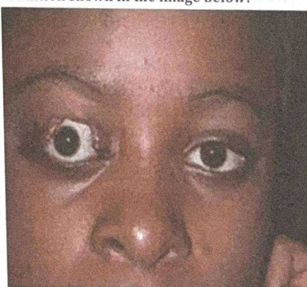
What is the most likely complication of the condition shown in the image below?

Which of the following intraocular foreign bodies causes severe toxicity and needs to be removed promptly?
Senile ptosis is:
Explanation: ***Modified Burrow's operation*** - The **Modified Burrow's operation** is primarily used in oculoplastic surgery for the correction of **entropion** or **ectropion**, and to resect redundant skin and muscle, not for ptosis correction. - It involves removing a strip of skin and orbicularis muscle, which does not address the underlying levator muscle weakness or disinsertion typical of congenital ptosis. *Blaskowics' operation* - This procedure involves an **anterior approach** to resect and advance the **levator aponeurosis** and Müller's muscle. - It is used for **mild to moderate congenital ptosis** with residual levator function (levator function >4-5 mm). *Everbusch's operation* - An **external approach** to resect the **levator muscle** or aponeurosis to elevate the eyelid. - It is used for **moderate congenital ptosis** where there is some residual levator function. *Frontalis sling operation* - This is a **suspension procedure** that connects the eyelid to the **frontalis muscle** using autogenous fascia lata, silicone rod, or other materials. - It is the procedure of choice for **severe congenital ptosis with poor levator function** (<4 mm), allowing the patient to use the frontalis muscle to elevate the eyelid.
Explanation: ***Kuhnt surgery*** - **Kuhnt surgery** refers to procedures for **ectropion**, which is the outward turning of the eyelid margin, not entropion. - This procedure typically involves **resection of the tarsus and conjunctiva** to tighten the lower lid and correct its eversion. *Wheeler Procedure* - The **Wheeler procedure** is a surgical technique used to correct **entropion**, particularly to address chronic or spastic forms. - It involves a skin and muscle flap to evert the eyelid margin. *Lateral tarsal split procedure* - The **lateral tarsal split procedure** is a common surgical method for **entropion** repair. - It involves a horizontal incision through the eyelid, often combined with placement of sutures, to evert the eyelid margin. *Quicke Procedure* - The **Quickert procedure** (often spelled "Quickert") is a surgical technique for **entropion** that involves everting sutures and sometimes a horizontal shortening of the eyelid. - It aims to reposition the eyelid margin to prevent the inward turning of the lashes.
Explanation: ***Symblepharon*** - **Symblepharon** is the term for the adhesion between the **palpebral conjunctiva** (lining the eyelid) and the **bulbar conjunctiva** (covering the eyeball). - This condition can limit eye movement and cause chronic irritation, often resulting from severe conjunctival inflammation or injury. *Trichiasis (inward growth of eyelashes)* - **Trichiasis** refers to the misdirection of eyelashes such that they rub against the cornea or conjunctiva. - It causes irritation, foreign body sensation, and can lead to corneal abrasion, but it does not involve fusion of conjunctival layers. *Ectropion (outward turning of eyelid)* - **Ectropion** is a condition where the lower eyelid turns outward or sags away from the eyeball. - This exposes the conjunctiva, causing dryness, irritation, and epiphora (excessive tearing), but it is not a fusion of conjunctival tissues. *Tylosis (thickening of skin on palms and soles)* - **Tylosis** is a medical term referring to diffuse **hyperkeratosis** or thickening of the skin, typically observed on the palms and soles. - This condition is completely unrelated to the conjunctiva or eye structures.
Explanation: ***Horner's syndrome*** - **Horner's syndrome** is characterized by **ptosis**, **miosis**, and **anhidrosis** on the affected side. - While it can manifest with a mild degree of **apparent enophthalmos**, this is primarily due to the **ptosis creating an illusion** of globe retraction and **not true enophthalmos**. - It is caused by disruption of the **sympathetic nervous supply**, not by actual posterior displacement of the globe. *Cicatricial changes* - **Cicatricial changes** (scarring) within the orbit can cause **traction on the globe**, pulling it inward and resulting in **true enophthalmos**. - This scarring can occur following **trauma**, **inflammation**, or **surgery** affecting the orbital tissues. *Orbital floor fracture* - An **orbital floor fracture** typically leads to **enophthalmos** due to **herniation of orbital contents** (fat, muscle) into the maxillary sinus. - This involves a **structural defect** with increased orbital volume and loss of support for the globe. *Loss of orbital fat* - **Loss of orbital fat**, often seen in conditions like **Romberg's disease**, severe dehydration, or aging, causes the globe to sink backward. - This is due to a **reduction in volume supporting the globe**, resulting in **true enophthalmos**.
Explanation: ***Abnormal extra row of cilia*** - **Distichiasis** is a congenital or acquired condition characterized by the presence of a double row of eyelashes, where the extra row emerges from the **Meibomian gland orifices**. - These accessory eyelashes can be the same length as normal lashes or appear finer and shorter, often causing **ocular irritation**, corneal abrasion, and epiphora due to their abnormal growth direction. *Abnormal inversion of eyelashes* - This description typically refers to **trichiasis**, where normally positioned eyelashes grow inwards towards the eye. - While both can cause irritation, **trichiasis** involves misdirection of existing lashes, whereas distichiasis involves an *extra* row. *Abnormal eversion of eyelashes* - Eversion of eyelashes is not a recognized abnormality in this context; rather, **ectropion** refers to the outward turning of the eyelid margin, which may expose the eyelashes but is not a primary cilial abnormality. - This condition is more about eyelid positioning than the eyelashes themselves. *Misdirected cilia* - While distichiasis does involve cilia growing in an abnormal direction, the key feature of distichiasis is the presence of an *additional* row of lashes, not just misdirection of the primary row. - **Trichiasis** is the more appropriate term for misdirected cilia from the normal lash line.
Explanation: ***Congenital ptosis*** - The **Fasanella-Servat operation** is indicated for **mild to moderate ptosis with good levator function** (levator function >10mm). - This includes cases of **mild congenital ptosis** where the levator muscle has adequate function. - The procedure involves resecting a portion of the **conjunctiva, Müller's muscle, and upper tarsus** to elevate the eyelid. - It provides approximately **2-3mm of lid elevation** and is particularly useful when levator function is preserved. *Horner syndrome* - Horner syndrome causes ptosis due to **denervation of Müller's muscle** (sympathetic dysfunction). - The Fasanella-Servat operation **resects Müller's muscle**, which would be counterproductive when this muscle is already dysfunctional. - Ptosis in Horner syndrome is typically managed with **levator resection** or observation, not Fasanella-Servat. *Myasthenia gravis* - Ocular manifestations of **myasthenia gravis** are treated with **acetylcholinesterase inhibitors** and immunomodulatory therapies. - The underlying **neuromuscular junction defect** causes variable ptosis that fluctuates throughout the day. - Surgical correction is not appropriate as the condition requires medical management of the autoimmune process. *Drug induced ptosis* - **Drug-induced ptosis** is a reversible condition that resolves with **discontinuation of the offending medication**. - Common culprits include topical prostaglandin analogs and certain systemic medications. - Surgical intervention like the Fasanella-Servat operation is not indicated as the cause is reversible.
Explanation: ***Bacterial origin*** - **Ligneous conjunctivitis** is primarily a genetic disorder due to **plasminogen deficiency**, not a bacterial infection. - While secondary bacterial infections can occur, they are not the underlying cause or hallmark feature of this condition. *Recurrent nature* - The disease is characterized by a **recurrent nature**, with pseudomembranes frequently reforming even after surgical removal. - This tendency for recurrence underscores the underlying systemic enzymatic deficiency. *Wood-like membranes* - The hallmark clinical feature of ligneous conjunctivitis is the formation of **dense, rigid, wood-like pseudomembranes** on the conjunctiva, particularly the palpebral conjunctiva. - These membranes are composed of fibrin and other extracellular matrix components, which accumulate due to impaired fibrinolysis. *Plasminogen deficiency* - Ligneous conjunctivitis is caused by a profound deficiency or dysfunction of **plasminogen**, an enzyme crucial for fibrinolysis. - This deficiency leads to impaired breakdown of fibrin, resulting in its abnormal accumulation in various tissues, most notably the eye.
Explanation: ***Exposure Keratitis*** - The image shows **proptosis** (exophthalmos) of the right eye, where the eyeball protrudes forward. This condition often leads to incomplete eyelid closure (lagophthalmos). - **Exposure keratitis** occurs when the cornea is inadequately covered by the eyelids, leading to drying and damage due to constant exposure to air and environmental factors. *Difficulty in eye movement* - While **proptosis** can sometimes be associated with restricted eye movements (e.g., in severe Graves' ophthalmopathy due to muscle swelling), it is not the **most likely direct complication** of the exposure itself. - The image primarily depicts the physical displacement of the globe, which predisposes to corneal issues, not necessarily oculomotor dysfunction as the primary complication. *Cataract* - **Cataracts** are opacities of the lens and are typically associated with aging, trauma, or certain systemic conditions (e.g., diabetes, steroid use). - They are not a direct or common complication of **proptosis** or the resulting **exposure of the ocular surface**. *Glaucoma* - **Glaucoma** is a group of conditions characterized by damage to the optic nerve, often due to elevated intraocular pressure. - While severe **proptosis** leading to orbital congestion can theoretically increase intraocular pressure, it is not the most direct or prevalent complication compared to **exposure keratitis**, which is a direct consequence of inadequate globe protection.
Explanation: ***Copper*** - **Copper foreign bodies** cause **chalcosis bulbi**, a severe toxic reaction characterized by: - **Greenish discoloration** of anterior lens capsule and cornea - **Retinal degeneration** and pigmentary changes - **Vision loss** if not removed promptly - **Pure copper is highly toxic** and mandates immediate surgical removal to prevent irreversible ocular damage *Aluminium* - **Aluminum foreign bodies** are **inert** and well-tolerated in the eye - Generally do not cause significant inflammatory reactions or toxicity - Removal not required unless causing mechanical complications *Chromium* - **Chromium foreign bodies** are **inert** and non-toxic - Do not cause significant inflammation or tissue damage - Removal typically not necessary unless mechanically disruptive *Nickel* - **Nickel foreign bodies** are **relatively inert** within the eye - Rarely cause toxic reactions or significant inflammation - Removal usually not required unless causing mechanical irritation
Explanation: **Explanation:** **Senile ptosis** (also known as Involutional ptosis) is the most common form of acquired ptosis in the elderly. **Why Aponeurotic is correct:** The primary pathology in senile ptosis is the **disinsertion, dehiscence, or stretching of the Levator Palpebrae Superioris (LPS) aponeurosis** from its attachment to the tarsal plate. This occurs due to age-related degenerative changes. Characteristically, these patients present with a **high or absent upper eyelid crease** and good levator function, as the muscle itself is healthy, but its "tendon" (aponeurosis) has slipped. **Why other options are incorrect:** * **Neurogenic:** Caused by nerve defects, such as 3rd Nerve Palsy or Horner’s Syndrome. Senile ptosis does not involve nerve dysfunction. * **Myogenic:** Caused by primary muscle disorders (e.g., Myasthenia Gravis or Myotonic Dystrophy). In senile ptosis, the LPS muscle fibers are typically normal. * **Mechanical:** Caused by the weight of a mass (tumor, edema, or chalazion) pulling the lid down. Senile ptosis is due to structural laxity, not added weight. **High-Yield Clinical Pearls for NEET-PG:** * **Clinical Sign:** A "high skin crease" is the hallmark of aponeurotic ptosis. * **Thinning of the lid:** The eyelid may appear thin, sometimes allowing the iris to be visible through the skin (due to LPS dehiscence). * **Surgical Management:** The treatment of choice is **LPS Aponeurosis advancement or repair**. * **Differential:** If ptosis is associated with miosis, think Horner’s; if associated with ocular motility issues, think 3rd Nerve Palsy.
Get full access to all questions, explanations, and performance tracking.
Start For Free