Which of the following ocular findings is not associated with diabetes?
What condition is suggested by eyelid papules and a hoarse cry in a child?
Unilateral frontal blisters with upper lid edema and conjunctivitis is seen in?
The tear drop sign on imaging is characteristically associated with:
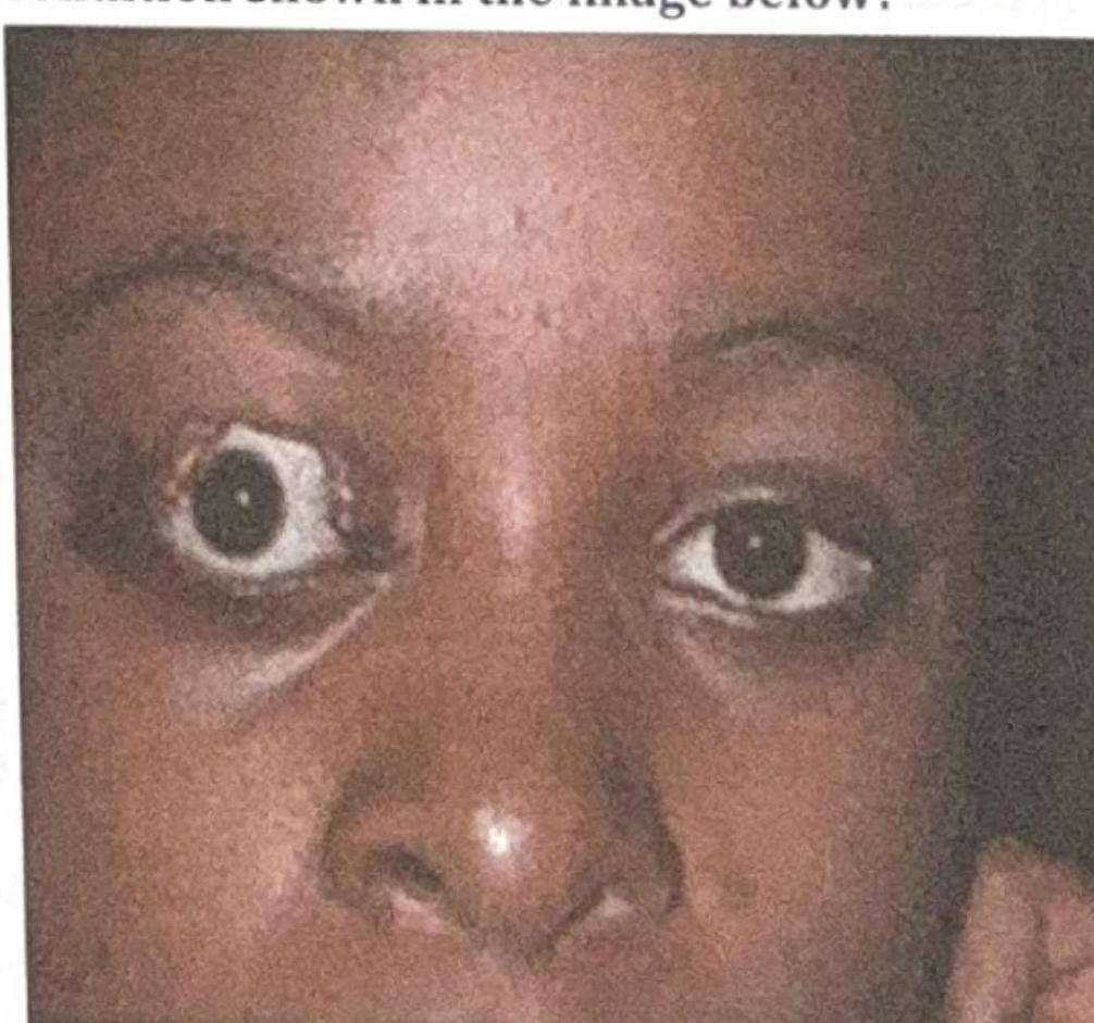
What is the most likely complication of the condition shown in the image below?

Distichiasis is a condition characterized by:
What is the term given to a condition with an extra layer of cilia posterior to grey line?
Chronic granulomatous inflammation in upper lid (painless swelling) is characteristic of:
Internal hordeolum is due to inflammation of-
What is the most common malignant tumour of eyelid?
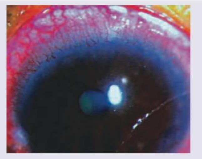
Explanation: ***Blepharophimosis*** - This is a **congenital disorder** characterized by small palpebral fissures, ptosis, and epicanthus inversus, which is **not associated with diabetes**. - It is a **developmental anomaly** of the eyelids, with no known link to metabolic conditions like diabetes. *Retinopathy* - **Diabetic retinopathy** is a common and serious complication of diabetes, caused by damage to the blood vessels in the retina. - It can lead to vision loss if not managed, and is directly linked to **poor glycemic control**. *Early senile cataract* - Diabetes is a significant risk factor for the **earlier development and progression of cataracts**, including senile cataracts. - High blood sugar levels can cause changes in the lens, leading to **opacification** and impaired vision. *Neovascular glaucoma* - This severe form of **secondary glaucoma** is often a complication of advanced **diabetic retinopathy**. - Ischemia in the retina triggers the growth of **new blood vessels** on the iris and in the angle of the eye, obstructing aqueous outflow and raising intraocular pressure.
Explanation: ***Lipoid proteinosis*** - This condition is characterized by **hoarseness from infancy** due to deposition in the vocal cords and characteristic **beaded papules on the eyelids** (moniliform blepharosis). - Also known as **Urbach-Wiethe disease**, it is a rare autosomal recessive disorder resulting from mutations in the **ECM1 gene**, leading to abnormal deposition of hyaline material in various tissues. *Croup* - Croup typically presents with a **barking cough** and **stridor**, often following a viral upper respiratory infection. - It does not cause eyelid papules or chronic hoarseness from infancy, but rather acute respiratory distress. *Acrodermatitis enteropathica* - This is a rare autosomal recessive disorder of **zinc malabsorption**, leading to a classic triad of **dermatitis**, **diarrhea**, and **alopecia**. - It does not involve eyelid papules or hoarseness as primary features. *Congenital syphilis* - Congenital syphilis can cause a wide range of manifestations, including skin rashes, bone abnormalities, and rhinitis ("snuffles"), but eyelid papules and chronic hoarseness are not typical presenting features. - Diagnosis is usually confirmed by serological tests for syphilis.
Explanation: ***Herpes Zoster Ophthalmicus*** - This condition is characterized by a **unilateral vesicular rash** (blisters) in the **trigeminal dermatome (V1)**, which includes the forehead and upper eyelid, along with significant **lid edema** and **conjunctivitis**. - **Hutchinson's sign** (lesions on the tip, side, or root of the nose) indicates a high risk of ocular involvement due to the nasociliary nerve innervation. *Acanthamoeba Keratitis* - This is an **amoebic infection** of the cornea typically associated with **contact lens wear** and often presents with severe pain and a **ring infiltrate** in the cornea. - It does not typically present with unilateral frontal blisters or significant lid edema. *Herpes Simplex* - Herpes simplex typically causes **recurrent corneal ulcers** (dendritic or geographic) and sometimes blepharitis, but not the widespread **unilateral frontal blisters** seen in the trigeminal distribution. - While it can cause conjunctivitis and lid edema, the pattern of skin lesions is the key differentiator. *Neuroparalytic Keratitis* - This condition results from **trigeminal nerve damage**, leading to corneal anesthesia and subsequent **trophic corneal ulceration**. - It presents primarily with **corneal findings** (epithelial defects, ulcers) due to impaired sensation and tear film stability, not initial vesicular skin lesions or prominent lid edema.
Explanation: ***floor of orbit*** - A **tear-drop sign** on imaging, particularly in X-rays or CT scans, is characteristic of a **blow-out fracture** of the orbit, specifically involving the **inferior orbital wall (floor)**. - The "tear-drop" appearance is created by the herniation of **orbital soft tissues (fat and/or inferior rectus muscle)** into the maxillary sinus through the fractured orbital floor. *Retinoblastoma* - Retinoblastoma is a **malignant tumor of the retina** in children, typically presenting with **leukocoria (white pupillary reflex)**, strabismus, or vision changes. - While imaging (CT or MRI) can show an intraocular mass with calcifications, it does not typically produce a "tear-drop sign." *Congenital nasolacrimal duct obstruction* - This condition presents with chronic **tearing (epiphora)** and discharge in infants due to failure of the nasolacrimal duct to open. - Imaging is usually not required for diagnosis, and when performed, it would not show a "tear-drop sign" but might reveal a dilated lacrimal sac. *Dry eyes* - Dry eyes (keratoconjunctivitis sicca) involve insufficient tear production or excessive tear evaporation, leading to ocular discomfort, burning, and foreign body sensation. - Diagnosis is clinical, involving tests like the **Schirmer test** or fluorescein staining, and it has no associated "tear-drop sign" on imaging.
Explanation: ***Exposure Keratitis*** - The image shows **proptosis** (exophthalmos) of the right eye, where the eyeball protrudes forward. This condition often leads to incomplete eyelid closure (lagophthalmos). - **Exposure keratitis** occurs when the cornea is inadequately covered by the eyelids, leading to drying and damage due to constant exposure to air and environmental factors. *Difficulty in eye movement* - While **proptosis** can sometimes be associated with restricted eye movements (e.g., in severe Graves' ophthalmopathy due to muscle swelling), it is not the **most likely direct complication** of the exposure itself. - The image primarily depicts the physical displacement of the globe, which predisposes to corneal issues, not necessarily oculomotor dysfunction as the primary complication. *Cataract* - **Cataracts** are opacities of the lens and are typically associated with aging, trauma, or certain systemic conditions (e.g., diabetes, steroid use). - They are not a direct or common complication of **proptosis** or the resulting **exposure of the ocular surface**. *Glaucoma* - **Glaucoma** is a group of conditions characterized by damage to the optic nerve, often due to elevated intraocular pressure. - While severe **proptosis** leading to orbital congestion can theoretically increase intraocular pressure, it is not the most direct or prevalent complication compared to **exposure keratitis**, which is a direct consequence of inadequate globe protection.
Explanation: ***Abnormal extra row of cilia*** - **Distichiasis** is a congenital or acquired condition characterized by the presence of a double row of eyelashes, where the extra row emerges from the **Meibomian gland orifices**. - These accessory eyelashes can be the same length as normal lashes or appear finer and shorter, often causing **ocular irritation**, corneal abrasion, and epiphora due to their abnormal growth direction. *Abnormal inversion of eyelashes* - This description typically refers to **trichiasis**, where normally positioned eyelashes grow inwards towards the eye. - While both can cause irritation, **trichiasis** involves misdirection of existing lashes, whereas distichiasis involves an *extra* row. *Abnormal eversion of eyelashes* - Eversion of eyelashes is not a recognized abnormality in this context; rather, **ectropion** refers to the outward turning of the eyelid margin, which may expose the eyelashes but is not a primary cilial abnormality. - This condition is more about eyelid positioning than the eyelashes themselves. *Misdirected cilia* - While distichiasis does involve cilia growing in an abnormal direction, the key feature of distichiasis is the presence of an *additional* row of lashes, not just misdirection of the primary row. - **Trichiasis** is the more appropriate term for misdirected cilia from the normal lash line.
Explanation: ***Distichiasis*** - This condition involves an **extra row of eyelashes** (cilia) that emerge from the **Meibomian gland orifices** on the posterior aspect of the eyelid margin, behind the normal lash line (posterior to the grey line). - These accessory lashes can be directed towards the globe, causing **corneal irritation** and damage. *Trichiasis* - This refers to the **misdirection of normally positioned eyelashes** towards the globe, causing irritation and corneal damage. - There is **no additional row of lashes** present, unlike in distichiasis. *Tylosis* - **Tylosis** is a thickening or **hyperkeratosis of the skin**, often seen in the soles of the feet and palms of the hands. - It does not refer to a condition related to eyelashes or the eyelid margin. *Madarosis* - **Madarosis** is the general term for **loss of eyelashes** (cilia) or eyebrows. - This is the opposite of having an extra layer of eyelashes.
Explanation: ***Chalazion*** - A chalazion is a **chronic**, sterile, **lipogranulomatous** inflammation of the **meibomian glands**. - It presents as a **painless**, firm, round swelling in the eyelid, often in the upper lid due to the larger meibomian glands. *Trachoma* - Trachoma is a **chronic keratoconjunctivitis** caused by *Chlamydia trachomatis*. - It primarily affects the conjunctiva and cornea, leading to scarring, entropion, and eventual blindness, not a painless eyelid swelling. *Internal Hordeolum* - An internal hordeolum is an **acute** bacterial infection of a **meibomian gland**, forming an abscess. - It is typically **painful**, red, and tender, contrasting with the painless nature of the given presentation. *External hordeolum* - An external hordeolum (stye) is an **acute** bacterial infection of the **glands of Zeis or Moll** at the lid margin. - It is usually **painful**, red, and tender, presenting as a small pustule or nodule on the eyelid margin, not a deep-seated painless swelling.
Explanation: ***Meibomian glands (Correct)*** - An **internal hordeolum** results from acute **bacterial infection** (usually *Staphylococcus aureus*) and inflammation of a **Meibomian gland**, which are modified sebaceous glands located within the tarsal plate of the eyelid. - These glands produce the **lipid layer** of the tear film, and their blockage and infection lead to a painful, red lump on the **inner surface of the eyelid**. *Moll's gland (Incorrect)* - **Moll's glands** are modified apocrine sweat glands located near the base of the eyelashes. - Inflammation or infection of a Moll's gland would more commonly contribute to an **external hordeolum (stye)**, not an internal one. *Lacrimal gland (Incorrect)* - The **lacrimal gland** produces the watery component of tears and is located in the superotemporal orbit. - Inflammation of the lacrimal gland is called **dacryoadenitis**, which presents with swelling in the outer part of the upper eyelid and is distinct from a hordeolum. *Zeis gland (Incorrect)* - **Zeis glands** are sebaceous glands associated with the hair follicles of the eyelashes. - Similar to Moll's glands, infection of a Zeis gland is a common cause of an **external hordeolum (stye)**, which appears on the eyelid margin.
Explanation: ***Basal cell cancer*** - **Basal cell carcinoma (BCC)** is by far the most common malignant tumor of the eyelid, accounting for approximately **90% of all eyelid malignancies**. - It typically appears as a **slow-growing nodule** with rolled borders, central ulceration, and telangiectasias, most commonly affecting the **lower eyelid and medial canthus**. - BCC is strongly associated with **chronic UV radiation exposure** and rarely metastasizes, but can cause significant local tissue destruction if untreated. *Sebaceous gland carcinoma* - **Sebaceous gland carcinoma** (also known as meibomian gland carcinoma) is a rare but aggressive malignant tumor arising from the sebaceous glands of the eyelid. - It accounts for approximately **1-5% of eyelid malignancies** and has a higher risk of metastasis compared to BCC. - More common in **Asian populations** and often masquerades as chronic blepharoconjunctivitis, leading to delayed diagnosis. *Squamous cell carcinoma* - **Squamous cell carcinoma (SCC)** is the **second most common** malignant eyelid tumor, accounting for approximately **5-10% of cases**. - It presents as a firm, erythematous nodule or plaque and has a higher metastatic potential than BCC. - Associated with **UV exposure, HPV infection**, and immunosuppression. *Melanoma* - **Melanoma** is a rare malignant tumor of the eyelid, accounting for less than **1% of eyelid malignancies**. - It arises from melanocytes and carries a significant risk of metastasis and mortality. - Presents as a pigmented lesion with irregular borders, but amelanotic variants can also occur.
Get full access to all questions, explanations, and performance tracking.
Start For Free