Which route is most preferred for Endophthalmitis treatment
Kayser-Fleischer rings (KF rings) are seen in:
A patient presents with acute appendicitis. What is NOT to be done?
Which of the following intraocular foreign bodies causes severe toxicity and needs to be removed promptly?
Perforating injuries with retained intraocular foreign body are more serious than those without because of:
Best method of detection of a retained glass intraocular foreign body is
In a patient with a metallic foreign body in the eye, which investigation should NOT be done?
A laborer working in a construction site has a history of chuna particles falling in eye. Next step of management is
Best method of detection of retained glass intraocular foreign body is:
Which of the following ophthalmologic conditions shows 'Vossius ring' during examination?
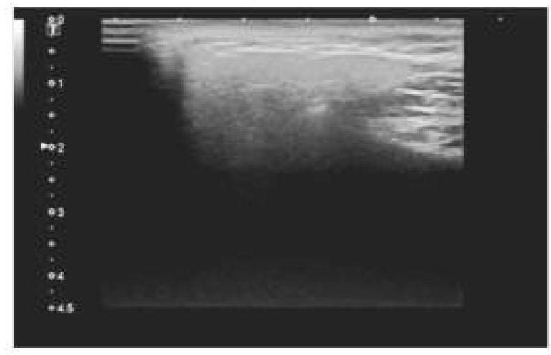
Explanation: ***Intravitreal antibiotic*** - **Intravitreal injection** ensures high concentrations of antibiotics directly reach the **vitreous cavity**, which is essential for treating intraocular infections like endophthalmitis effectively. - This route bypasses ocular barriers, achieving therapeutic levels at the site of infection that would be difficult to attain with systemic or topical approaches. *Topical antibiotic* - **Topical antibiotics** have limited penetration into the **vitreous**, making them generally ineffective as a sole therapy for established endophthalmitis. - They are primarily used for **conjunctivitis** or prophylaxis after surgery, not for deep-seated intraocular infections. *Intravenous antibiotic* - While **intravenous antibiotics** can achieve systemic levels, their ability to cross the **blood-retinal barrier** and reach effective concentrations in the vitreous is often insufficient for endophthalmitis. - They may be used as an **adjunct therapy** but are not preferred as the primary route for direct infection treatment. *Oral antibiotic* - **Oral antibiotics** have poor penetration into the **vitreous cavity**, similar to intravenous drugs, and are generally inadequate for treating endophthalmitis. - They are not considered a primary treatment route due to the rapid progression and potential for severe vision loss associated with the condition.
Explanation: ***Wilson's disease*** - **Kayser-Fleischer (KF) rings** are pathognomonic for **Wilson's disease**, resulting from **copper deposition** in the Descemet's membrane of the cornea. - This genetic disorder leads to excessive **copper accumulation** in the liver, brain, and other tissues due to impaired copper excretion. *Pterygium* - A **pterygium** is a benign growth of the **conjunctiva** that extends onto the cornea, typically appearing as a fleshy, triangular lesion. - It is not associated with systemic copper metabolism disorders or the presence of KF rings. *Hemochromatosis* - **Hemochromatosis** is a disorder of **iron overload**, leading to iron deposition in various organs, including the liver, heart, and pancreas. - It does not involve copper metabolism or the formation of corneal rings. *Menke's kinked hair syndrome* - **Menke's syndrome** is a genetic disorder characterized by **copper deficiency**, leading to impaired copper transport and utilization. - It presents with severe neurological deterioration, connective tissue abnormalities, and characteristic **kinky hair**, but not KF rings.
Explanation: ***Check for visual acuity*** - **Visual acuity** assessment is not relevant to the diagnosis or management of **acute appendicitis**. - This examination is typically performed in cases of suspected eye injury, vision changes, or neurological issues that affect vision. - In the context of acute appendicitis, checking visual acuity would be inappropriate and waste valuable time. *Give antibiotics* - **Antibiotics** are crucial in managing **acute appendicitis** to prevent progression to perforation and reduce postoperative infection risk. - They are typically administered preoperatively and continued postoperatively, especially in cases of complicated appendicitis. - Broad-spectrum antibiotics covering **gram-negative organisms and anaerobes** are standard practice. *Do primary survey* - A **primary survey** (ABCDE approach) is essential in any emergent patient presentation to assess and manage immediate **life-threatening conditions**. - While appendicitis itself may not be immediately life-threatening, ensuring patient stability and ruling out other serious conditions is critical. - This is standard emergency medicine practice and should always be performed. *Perform appendectomy* - **Appendectomy** (surgical removal of the appendix) is the definitive treatment for **acute appendicitis**. - This is the standard of care and should be performed once the diagnosis is confirmed and the patient is stable. - Either open or laparoscopic approach can be used depending on clinical factors and surgeon expertise.
Explanation: ***Copper*** - **Copper foreign bodies** cause **chalcosis bulbi**, a severe toxic reaction characterized by: - **Greenish discoloration** of anterior lens capsule and cornea - **Retinal degeneration** and pigmentary changes - **Vision loss** if not removed promptly - **Pure copper is highly toxic** and mandates immediate surgical removal to prevent irreversible ocular damage *Aluminium* - **Aluminum foreign bodies** are **inert** and well-tolerated in the eye - Generally do not cause significant inflammatory reactions or toxicity - Removal not required unless causing mechanical complications *Chromium* - **Chromium foreign bodies** are **inert** and non-toxic - Do not cause significant inflammation or tissue damage - Removal typically not necessary unless mechanically disruptive *Nickel* - **Nickel foreign bodies** are **relatively inert** within the eye - Rarely cause toxic reactions or significant inflammation - Removal usually not required unless causing mechanical irritation
Explanation: ***Deleterious effects of foreign bodies*** - This is the **MOST SPECIFIC and PRIMARY reason** that distinguishes retained IOFBs from perforating injuries without retained foreign bodies. - Retained intraocular foreign bodies cause **direct toxic effects** on ocular tissues depending on their composition: **siderosis bulbi** from iron (causing rust-colored deposits, retinal degeneration, and vision loss), **chalcosis** from copper (greenish deposits and inflammation), and direct mechanical trauma to delicate intraocular structures. - These **material-specific toxic effects** are unique to retained foreign bodies and occur regardless of whether infection or inflammation develops. - The foreign body acts as a constant source of **chronic inflammation and tissue damage**, leading to complications like cataract, glaucoma, retinal detachment, and progressive vision loss. *More chances of infection* - While retained IOFBs do increase the risk of **endophthalmitis** (severe intraocular infection), infection risk exists with any perforating injury, whether or not a foreign body is retained. - The question asks what makes retained IOFB cases **MORE serious** - the infection risk is elevated but not the PRIMARY distinguishing feature. - Prophylactic antibiotics can reduce infection risk, but cannot prevent the direct toxic effects of the retained material. *More chances of sympathetic ophthalmitis* - Sympathetic ophthalmitis is a rare bilateral granulomatous uveitis that can occur after **penetrating ocular trauma with uveal tissue injury**. - This risk exists with perforating injuries in general, not specifically because of the retained foreign body itself. - The presence of a foreign body is less important than uveal prolapse and inflammation in triggering this immune-mediated response. *All of the options* - While infection and sympathetic ophthalmitis are legitimate concerns, they are **not specific to retained foreign bodies** - they can occur with any penetrating injury. - The **direct deleterious/toxic effects** of the foreign body material (siderosis, chalcosis, mechanical damage) are the PRIMARY and MOST SPECIFIC reason that makes retained IOFB cases more serious. - This option is incorrect because it doesn't distinguish the unique hazard posed by the retained foreign body itself.
Explanation: ***CT scan*** - **CT scans** are highly sensitive and specific for detecting **retained glass intraocular foreign bodies** due to their excellent spatial resolution and ability to visualize foreign materials with different densities. - Unlike MRI, CT is safe with metallic foreign bodies and provides precise localization, aiding surgical planning. *Radiography* - While helpful for detecting radio-opaque foreign bodies like metal, **plain X-rays display limited soft tissue contrast** and may struggle to visualize small or less dense objects like glass effectively within the complex orbital structures. - **Glass foreign bodies can be difficult to discern** from surrounding bony structures or soft tissues on conventional radiographs, leading to false negatives. *USG* - **Ultrasound (USG)** is effective for imaging soft tissues and can detect some foreign bodies, but its utility is limited when the object is small, non-reflective, or located deep within the globe, especially behind structures like the lens or iris. - **Acoustic shadowing** and artifact creation can also obscure the foreign body or mimic its presence, reducing diagnostic accuracy for glass. *Tonometry* - **Tonometry measures intraocular pressure** and is primarily used to screen for or monitor glaucoma. - It provides no information about the presence or location of **intraocular foreign bodies**.
Explanation: ***MRI*** - **Magnetic Resonance Imaging (MRI)** is contraindicated in patients with suspected **metallic foreign bodies** in the eye. - The powerful magnetic fields of an MRI can cause the metallic object to move, potentially leading to further **tissue damage** or even loss of vision. *X-ray* - **X-rays** are often the initial investigation of choice for detecting **radio-opaque foreign bodies** within the eye. - They can effectively localize larger metallic objects and are readily available in most emergency settings. *CT* - **Computed Tomography (CT)** scans provide detailed cross-sectional images and are excellent for precisely localizing **intraocular foreign bodies**, especially smaller ones. - CT can differentiate between metallic and non-metallic objects and assess for associated injuries like orbital fractures. *USG* - **Ultrasound (USG)** of the eye can be useful for detecting **intraocular foreign bodies**, especially if they are non-metallic or located in the posterior segment. - It can also assess for associated complications such as **retinal detachment** or vitreous hemorrhage.
Explanation: ***Irrigation with normal saline*** - The immediate priority for **chemical eye injuries**, such as those from **chuna (calcium hydroxide)**, is copious and prolonged irrigation to neutralize or dilute the causative agent and prevent further tissue damage. - **Alkali burns** are particularly dangerous as they can continue to penetrate deeper tissues, making immediate and thorough irrigation with neutral solutions like normal saline crucial. *Topical antibiotics* - While antibiotics might be used later to prevent secondary infections, they are **not the immediate next step** in managing an acute chemical burn. - Ignoring immediate irrigation increases the risk of severe damage, even if antibiotics are applied. *Padding the eye and analgesics* - **Padding the eye** can trap the chemical and prevent it from being effectively diluted or washed away, potentially worsening the injury. - **Analgesics** address pain but do not treat the underlying chemical injury or prevent ongoing damage to ocular tissues. *Complete scraping* - **Scraping** should generally be avoided in the acute phase of a chemical burn as it can cause further mechanical damage to an already compromised cornea and conjunctiva. - The primary goal is chemical neutralization and dilution through irrigation.
Explanation: ***CT scan*** - **CT scans** are highly sensitive for detecting **intraocular foreign bodies**, especially radiopaque materials like glass, and can accurately localize them within the eye. - They provide detailed cross-sectional images, which are crucial for surgical planning and assessing associated orbital injuries. - CT is particularly useful when **ultrasound is contraindicated** (suspected globe rupture) or for **metallic foreign bodies**. *Ultrasonography* - **Ultrasonography (B-scan)** is highly effective for detecting glass foreign bodies, as glass creates **high reflectivity** on ultrasound imaging. - It provides excellent visualization of intraocular structures and can detect both radiopaque and radiolucent foreign bodies. - However, it is **contraindicated** if globe rupture is suspected due to the risk of extruding intraocular contents, making CT the safer alternative in such cases. - Its utility can be limited by acoustic shadowing and requires skilled interpretation. *Radiography* - While **radiography** can detect some metallic foreign bodies, it has **limited sensitivity** for small or non-metallic intraocular foreign bodies like glass due to potential superimposition of bony structures. - Its two-dimensional nature can also make precise **localization difficult**. *Tonography* - **Tonography** is a diagnostic test used to measure the **outflow of aqueous humor** from the eye, primarily to evaluate for glaucoma. - It is **not used for detecting or localizing foreign bodies**; its purpose is entirely different, focusing on intraocular pressure dynamics.
Explanation: ***Traumatic iritis*** - A **Vossius ring** is a circular pigment deposit on the anterior lens capsule, formed by the impact of the iris during **ocular trauma**. - Its presence is a clear indicator of **blunt globe trauma**, which often leads to traumatic iritis. *Angle closure glaucoma* - This condition involves an **acute increase in intraocular pressure** due to the iris blocking the drainage angle. - While it can cause severe pain and vision loss, it does not involve the formation of a **Vossius ring**. *Retinal detachment* - This condition involves the separation of the sensory retina from the underlying retinal pigment epithelium. - Symptoms include **flashing lights**, **floaters**, and a **"curtain" vision loss**, with no association with a Vossius ring. *Corneal ulcer* - A corneal ulcer is an **open sore on the cornea**, typically caused by infection or injury. - It presents with **pain**, **photophobia**, **redness**, and **discharge**, without the characteristic lens pigment deposit.
Get full access to all questions, explanations, and performance tracking.
Start For Free