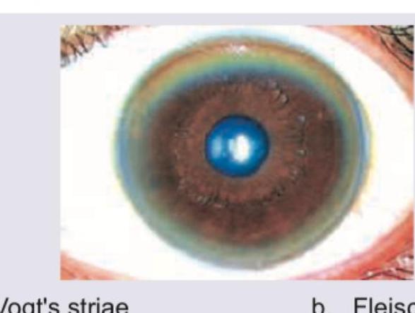
A patient presents with the clinical findings shown in the image. What is the most likely diagnosis?

A 15-year-old child was diagnosed with Fanconi's syndrome. His school grades have been consistently poor and he has involuntary movements in his hands. Drug screening is negative. Eye finding shows: (APPG 2016)

Of the following ocular manifestations of Vitamin A deficiency, the first sign that can be clinically seen is:
The most common ocular lesion peculiar to HIV infection in early stage is :
In patients with systemic hypertension, the characteristic haemorrhage seen on ophthalmoscopy is
Consider the following : 1. Night blindness 2. Corneal xerosis 3. Conjunctival xerosis 4. Keratomalacia What is the correct sequence of the above in the progress of clinical presentation of vitamin A deficiency blindness?
All of the following are false about eye lid signs in GRAVES DISEASE except?
Sarcoidosis is associated with
A 12-year-old boy is admitted to the emergency department with signs of meningitis. To determine the specific type of meningitis, it is necessary to aspirate cerebrospinal fluid with a lumbar puncture for laboratory examination. However, before performing a lumbar puncture, it must be established that the cerebrospinal fluid pressure is not elevated. What condition in the eye would indicate that cerebrospinal fluid pressure is too elevated for a lumbar puncture to be performed?
All are manifestation of dengue virus infection in eye except?
Explanation: ***Herpes zoster ophthalmicus*** - The image shows a classic vesicular rash following the **dermatomal distribution** of the trigeminal nerve (V1 branch), affecting the forehead, eyelid, and bridge of the nose. - The involvement of the eye and surrounding tissue, particularly with vesicles on the tip of the nose (**Hutchinson's sign**), indicates potential ocular involvement and confirms Herpes zoster ophthalmicus. *Orbital cellulitis* - Characterized by **painful proptosis**, **ophthalmoplegia**, and **vision loss**, which are not clearly visible or suggested by the skin lesions. - While there is periorbital swelling, the distinctive vesicular rash in a dermatomal pattern is absent in orbital cellulitis. *Angle closure glaucoma* - Presents with a **sudden onset of severe eye pain**, blurred vision, **fixed mid-dilated pupil**, and colored halos, without external skin lesions. - This condition is an intraocular emergency and does not involve the characteristic rash seen in the image. *Subcutaneous emphysema* - Involves **air trapped under the skin**, causing a characteristic **crepitus** on palpation. - It often results from trauma or surgery and does not present with vesicular skin lesions.
Explanation: ***KF ring*** - The combination of **Fanconi's syndrome** (renal tubular acidosis), **poor school grades** (neurological deterioration), and **involuntary movements** (extrapyramidal symptoms) is highly suggestive of **Wilson's disease**. - **Kayser-Fleischer (KF) rings** are characteristic ocular findings in Wilson's disease, caused by **copper deposition** in the Descemet membrane of the cornea, which appears as a greenish-brown ring. *Vogt's striae* - **Vogt's striae** are vertical stress lines in the deep stroma and Descemet's membrane of the cornea, typically seen in advanced **keratoconus**. - While keratoconus can be associated with certain syndromes, it is not directly linked to the neurological and renal symptoms described in the patient. *Fleischer ring* - A **Fleischer ring** is a deposition of **iron pigment (hemosiderin)** in the basal epithelial cells of the cornea, often seen at the base of the cone in **keratoconus**. - Like Vogt's striae, it is an indicator of keratoconus and does not align with the systemic symptoms presented. *Stocker line* - A **Stocker line** is a **ferric oxide deposition** that appears as a brown line on the corneal epithelium, typically associated with the advancing edge of a **pterygium**. - This finding is specific to pterygium and is not systemic, thus not explaining the patient's neurological and renal issues.
Explanation: ***Conjunctival Xerosis*** - This is the **earliest clinical sign** of vitamin A deficiency in the eye, characterized by dryness of the conjunctiva. - It often progresses from a dull, lusterless appearance to the development of **Bitot's spots**. *Bitot's spots* - These are **foamy, triangular patches** of keratinized epithelium on the conjunctiva, usually temporal to the cornea. - While a prominent sign, they appear **after** the initial dryness of conjunctival xerosis. *Nyctalopia* - Also known as **night blindness**, this is a **functional symptom** rather than a visible clinical sign. - It indicates impaired vision in low light conditions due to dysfunction of the **retinal rods**, which is a consequence of vitamin A deficiency. *Corneal Xerosis* - This is a more **advanced stage** of vitamin A deficiency, where the dryness has progressed to affect the cornea. - It appears **after** conjunctival xerosis and can lead to more severe complications like corneal ulceration and keratomalacia.
Explanation: ***Soft exudates in retina*** - **Soft exudates**, also known as **cotton wool spots**, are the most common early ocular manifestation in HIV infection. - They represent ischemia of the nerve fiber layer due to microvascular occlusion and do not generally affect vision significantly. *Retinal necroses* - **Retinal necroses**, such as those seen in **progressive outer retinal necrosis (PORN)** or **acute retinal necrosis (ARN)**, are typically severe and rapidly progressive. - They are usually associated with advanced HIV infection (low CD4 count) and viral etiologies like **CMV retinitis** or **VZV**, not early stage HIV. *Retinal hemorrhages* - **Retinal hemorrhages** can occur in HIV, but they are often associated with systemic conditions like **thrombocytopenia** or **anemia**, or advanced opportunistic infections. - They are not considered the *most common* lesion peculiar to early HIV itself. *Kaposi's sarcoma* - **Kaposi's sarcoma** is a vascular tumor caused by **HHV-8 infection** and is associated with advanced immunosuppression in HIV. - While it can affect the conjunctiva or eyelids, primary intraocular involvement is rare and typically presents in later stages, not early HIV.
Explanation: ***Flame-shaped haemorrhage*** - **Flame-shaped hemorrhages** are superficial, linear hemorrhages occurring in the **nerve fiber layer** of the retina, characteristic of **hypertensive retinopathy**. - They result from the rupture of superficial capillaries due to **elevated intravascular pressure** in systemic hypertension. *Wet sponge haemorrhage* - This term is not a standard description for retinal hemorrhage types. - Retinal hemorrhages are typically categorized by their shape and depth, such as dot-and-blot, flame-shaped, or subretinal. *Suprachoroidal haemorrhage* - **Suprachoroidal hemorrhages** occur between the choroid and the sclera, often due to trauma or surgical complications. - They are much deeper than retinal hemorrhages and are typically not a direct, primary manifestation of chronic systemic hypertension on routine ophthalmoscopy. *Dot haemorrhage* - **Dot haemorrhages** are deep, punctate hemorrhages occurring in the **inner nuclear** or **outer plexiform layers** of the retina. - While they can be seen in various conditions including diabetic retinopathy, they are not the most characteristic type of hemorrhage seen specifically in systemic hypertension.
Explanation: ***1-3-2-4*** - This is the **correct sequence** according to WHO classification of xerophthalmia in vitamin A deficiency. - **Night blindness (XN)** is typically the **earliest clinical manifestation**, resulting from impaired rod photoreceptor function due to insufficient rhodopsin synthesis. - This is followed by **conjunctival xerosis (X1A)**, characterized by dryness and thickening of the bulbar conjunctiva, often accompanied by Bitot's spots (X1B) - foamy triangular patches of keratinized epithelium. - As deficiency progresses, **corneal xerosis (X2)** develops, with dryness extending to the corneal surface, creating a hazy, lusterless appearance. - The final and most severe stage is **keratomalacia (X3)**, involving corneal softening, ulceration, and potential perforation leading to irreversible blindness. *2-1-4-3* - This sequence incorrectly places **corneal xerosis** before **night blindness**. - Night blindness is a functional symptom that appears early, while corneal xerosis is a later structural change. - The progression also illogically places keratomalacia before conjunctival xerosis. *1-2-3-4* - This sequence incorrectly suggests **corneal xerosis** precedes **conjunctival xerosis**. - In vitamin A deficiency, xerosis (dryness) affects the **conjunctiva first**, then progresses to involve the **cornea**. - The conjunctiva is more susceptible to early changes than the cornea. *1-3-4-2* - This sequence incorrectly places **keratomalacia** before **corneal xerosis**. - Keratomalacia represents corneal softening and melting, which cannot occur before the cornea becomes xerotic (dry). - Corneal xerosis is a **prerequisite** for the development of keratomalacia.
Explanation: **⚠️ QUESTION ISSUE: All four options are TRUE statements about Graves' disease, making this question technically flawed.** ***Stellwag's sign: Incomplete and infrequent blinking*** ✓ - **Stellwag's sign** is correctly defined as **incomplete and infrequent blinking**, causing a characteristic staring appearance. - This is a **TRUE** sign of **Graves' ophthalmopathy** due to sympathetic overactivity. - **Marked as correct answer**, but all options are actually true. *Mobius sign: convergence insufficiency* ✓ - **Moebius sign** is correctly defined as **convergence insufficiency** (inability to converge eyes for near vision). - This is a **TRUE** sign of **Graves' disease**. - Statement is medically accurate. *Dalrymple sign: lid retraction* ✓ - **Dalrymple's sign** is correctly defined as **lid retraction** (upper eyelid retraction exposing sclera above iris). - This is a **TRUE** and classic sign of **Graves' ophthalmopathy**. - Statement is medically accurate. *Von Graefe's sign: lid lag on downgaze* ✓ - **Von Graefe's sign** is correctly defined as **lid lag on downgaze** (upper eyelid lags behind eyeball during downward gaze). - This is a **TRUE** sign of **Graves' disease**. - Statement is medically accurate. **Educational Note:** This FMGE-2017 question is problematic because all four eyelid sign descriptions are medically accurate. In the actual exam, Stellwag's sign was likely considered the "best" answer, possibly because it's the most specific or commonly tested.
Explanation: ***Anterior uveitis*** - **Anterior uveitis**, particularly chronic granulomatous anterior uveitis, is the most common ocular manifestation of **sarcoidosis**. - It results from **non-caseating granulomas** affecting the iris and ciliary body. - Sarcoidosis can also cause intermediate uveitis, posterior uveitis, and panuveitis, but anterior uveitis is most frequently seen. - Characteristic findings include mutton-fat keratic precipitates and iris nodules (Koeppe and Busacca nodules). *Keratitis* - **Keratitis** (inflammation of the cornea) is less commonly associated with sarcoidosis compared to uveitis. - While ocular sarcoidosis can rarely cause interstitial keratitis or band keratopathy, direct corneal inflammation is not a hallmark feature. *Cataract* - **Cataracts** are not directly caused by sarcoidosis itself, but can be a complication of chronic uveitis or prolonged corticosteroid treatment. - The disease process of sarcoidosis does not primarily involve cataract formation. *Ectopia lentis* - **Ectopia lentis** (lens dislocation) is a rare condition usually associated with genetic disorders like **Marfan syndrome** or homocystinuria. - It is not a recognized ocular manifestation of sarcoidosis.
Explanation: ***Papilledema*** - **Papilledema** is **swelling of the optic disc** due to increased intracranial pressure (ICP), which can be visualized during an **ophthalmoscopic examination**. - Performing a **lumbar puncture (LP)** in the presence of papilledema can lead to **brain herniation** due to a sudden drop in pressure below the spinal cord, creating a pressure gradient that forces brain tissue downward. - This is the **primary ocular contraindication** to LP and must be assessed before the procedure. *Retinal hemorrhages at the fovea* - **Retinal hemorrhages** at the fovea are not a direct sign of **increased intracranial pressure (ICP)** in the same way papilledema is. - While certain conditions causing elevated ICP can lead to retinal hemorrhages (e.g., severe hypertension, Terson syndrome), they are not the primary or most reliable indicator for contraindicating an **LP** compared to papilledema. - Retinal hemorrhages can occur from various causes including diabetic retinopathy, retinal vein occlusion, or trauma. *Obvious opacity of the lens* - An **obvious opacity of the lens** refers to a **cataract**. - **Cataracts** are lens opacities that impair vision and are typically associated with aging, trauma, or systemic diseases like diabetes, but not directly with **elevated CSF pressure** or as a contraindication for **LP**. *Separation of the pars optica retinae anterior to the ora serrata* - The **pars optica retinae** refers to the photosensitive posterior part of the retina, and the **ora serrata** is its anterior boundary. - **Separation** in this area might suggest a **retinal detachment** or other structural retinal issue, which is not an indicator of **elevated intracranial pressure (ICP)** and would not contraindicate a **lumbar puncture**.
Explanation: ***Cataract*** - **Cataracts** are primarily associated with aging, congenital factors, trauma, or long-term steroid use, not directly with acute dengue virus infection. - While dengue can cause various ocular manifestations, the formation of cataracts is a **chronic process** that does not fit the typical acute or subacute presentation of dengue-related eye complications. *Vitreous hemorrhage* - **Vitreous hemorrhage** can occur in dengue due to associated **thrombocytopenia** and coagulation abnormalities, leading to bleeding in the eye. - Severe dengue can induce systemic vasculopathy and bleeding tendencies, which may manifest as intraocular hemorrhage. *Maculopathy* - **Dengue maculopathy** is a recognized complication, often presenting as macular edema, hemorrhage, or foveolitis, leading to visual impairment. - This is thought to be due to direct viral effects, immune-mediated responses, or vasculitis affecting the retinal microvasculature. *Optic neuritis* - **Optic neuritis** following dengue infection has been reported, characterized by inflammation of the optic nerve, causing acute vision loss. - This is considered to be an **immune-mediated post-infectious complication** rather than a direct viral cytopathic effect on the nerve.
Diabetes Mellitus
Practice Questions
Hypertension
Practice Questions
Autoimmune Disorders
Practice Questions
Thyroid Disease
Practice Questions
HIV and AIDS
Practice Questions
Hematological Disorders
Practice Questions
Neurological Disorders
Practice Questions
Dermatological Conditions
Practice Questions
Pregnancy-Related Eye Changes
Practice Questions
Metabolic Disorders
Practice Questions
Ocular Toxicity of Systemic Medications
Practice Questions
Infectious Systemic Diseases
Practice Questions
Get full access to all questions, explanations, and performance tracking.
Start For Free