River blindness is caused by which of the following?
Essential atrophy of the choroid is due to an inborn error of metabolism of which amino acid?
All of the following are ocular symptoms seen in Herpes ophthalmicus, EXCEPT:
Uveitis is most commonly associated with which condition?
What condition is characterized by "candle-wax spots" in the retina?
Conjunctival xerosis is caused by which vitamin deficiency?
Optic nerve glioma is seen in?
Which vitamin deficiency causes xerophthalmia, a condition which causes dry eyes?
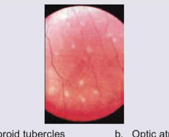
What does the fundus of this immunocompromised patient show?

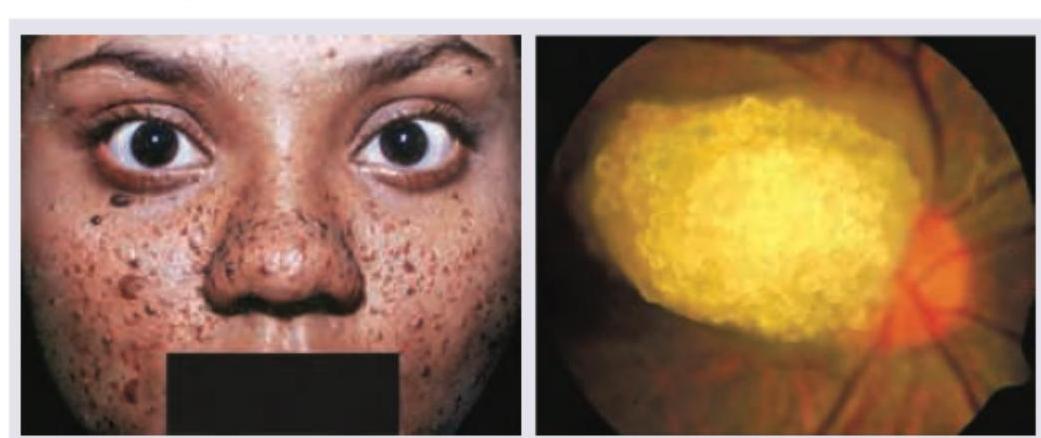
A 16-year-old girl with history of epilepsy has the presentation as shown below. Select the correct option:

Explanation: **Explanation:** **River Blindness**, medically known as **Onchocerciasis**, is caused by the nematode parasite **Onchocerca volvulus**. It is transmitted to humans through the bite of an infected **Blackfly (Simulium species)**, which breeds in fast-flowing rivers and streams—hence the name "River Blindness." 1. **Why Option B is Correct:** Once the larvae enter the body, they mature into adult worms in subcutaneous nodules. These adults release millions of **microfilariae** that migrate to the skin and eyes. In the eye, they cause severe inflammatory reactions leading to **punctate keratitis** ("snowflake" opacities), sclerosing keratitis, uveitis, and optic atrophy, eventually resulting in irreversible blindness. 2. **Why Other Options are Incorrect:** * **Option A:** While many parasitic infections are water-borne, Onchocerciasis is vector-borne (Blackfly) and not acquired by ingestion. * **Option C:** *Toxocara canis* (not Toxoplasma) causes **Ocular Larva Migrans**, typically presenting as a posterior pole granuloma or endophthalmitis in children. * **Option D:** Glaucoma is a group of eye conditions that damage the optic nerve, often due to high intraocular pressure, but it is not the causative agent of River Blindness. **High-Yield Clinical Pearls for NEET-PG:** * **Vector:** *Simulium* (Blackfly). * **Drug of Choice:** **Ivermectin** (kills microfilariae but not adult worms). * **Diagnosis:** "Skin snip" preparation to identify microfilariae or visualization of microfilariae in the anterior chamber using a slit lamp. * **Mazzotti Reaction:** A severe systemic reaction (fever, rash, hypotension) that can occur after treatment with Diethylcarbamazine (DEC) due to rapid microfilarial death; hence, DEC is generally avoided.
Explanation: **Explanation:** **Gyrate Atrophy of the Choroid and Retina** (also known as essential atrophy of the choroid) is an autosomal recessive dystrophy caused by a deficiency in the mitochondrial enzyme **ornithine aminotransferase (OAT)**. This enzyme is responsible for converting ornithine into glutamate and proline. A deficiency leads to a 10- to 20-fold increase in plasma **ornithine** levels (hyperornithinemia), which is toxic to the retinal pigment epithelium (RPE) and choroid. **Why the other options are incorrect:** * **Cystine & Cysteine:** These are sulfur-containing amino acids. Their metabolic derangement is associated with **Cystinosis**, which presents with pathognomonic needle-shaped crystals in the cornea and conjunctiva, not primary chorioretinal atrophy. * **Arginine:** While ornithine is a metabolic product of arginine (via the urea cycle), the primary defect in Gyrate Atrophy specifically involves the inability to process ornithine. **High-Yield Clinical Pearls for NEET-PG:** * **Clinical Presentation:** Patients typically present in the first decade with night blindness (nyctalopia) and constricted visual fields. * **Fundus Appearance:** Characteristic "scalloped," well-defined areas of chorioretinal atrophy starting in the mid-periphery and progressing centrally. * **Associated Features:** High myopia, posterior subcapsular cataracts, and cystoid macular edema (CME). * **Treatment:** A **Vitamin B6 (Pyridoxine)** trial is essential, as a subset of patients are "B6-responsive" and show a reduction in ornithine levels. Dietary restriction of arginine is also recommended.
Explanation: **Explanation:** **Herpes Zoster Ophthalmicus (HZO)** is caused by the reactivation of the Varicella-Zoster virus in the ophthalmic division of the trigeminal nerve ($V_1$). **Why Option A is the Correct Answer:** Oculomotor nerve involvement is a **sign**, not a **symptom**. In medical terminology, symptoms are subjective experiences reported by the patient (e.g., pain, blurred vision), whereas nerve palsies are objective clinical signs observed by the physician. While HZO can occasionally cause cranial nerve palsies (III, IV, or VI) due to contiguous inflammation, these are categorized as neurological complications/signs rather than primary ocular symptoms. **Analysis of Incorrect Options:** * **B. Disciform Keratitis:** This is a common corneal manifestation of HZO. It is an immune-mediated endotheliitis characterized by a central, disc-shaped area of stromal edema. * **C. Anterior Uveitis:** HZO frequently causes a high-pressure, non-granulomatous anterior uveitis. It is often associated with sectorial iris atrophy due to vasculitis. * **D. Nummular Keratitis:** This is a classic feature of HZO, presenting as multiple, small, granular subepithelial opacities (resembling coins) in the corneal stroma. **High-Yield Clinical Pearls for NEET-PG:** * **Hutchinson’s Sign:** Vesicles on the tip or side of the nose indicate involvement of the nasociliary nerve, which strongly predicts intraocular involvement. * **Pseudodendrites:** Unlike the true dendrites of Herpes Simplex (which have terminal bulbs and stain with fluorescein), HZO produces "pseudodendrites" which are elevated, lack terminal bulbs, and stain poorly. * **Treatment:** Oral Acyclovir (800 mg 5 times daily for 7–10 days) is the gold standard, ideally started within 72 hours of rash onset.
Explanation: **Explanation:** The association between systemic inflammatory disorders and the eye is a high-yield topic in Ophthalmology. Among the pediatric arthritides, **Pauciarticular (Oligoarticular) Juvenile Rheumatoid Arthritis (JRA)**—now more commonly termed Juvenile Idiopathic Arthritis (JIA)—is the condition most frequently associated with uveitis. **1. Why Pauciarticular JRA is Correct:** Pauciarticular JRA involves four or fewer joints during the first six months of the disease. Approximately **20-30%** of these patients develop chronic, non-granulomatous anterior uveitis. The risk is highest in young girls who are **ANA (Antinuclear Antibody) positive**. Crucially, the uveitis is often "white and quiet" (asymptomatic), making regular screening essential to prevent complications like band-shaped keratopathy and cataracts. **2. Why the Other Options are Incorrect:** * **Rheumatoid Arthritis (RA):** In adults, RA is more commonly associated with **Scleritis and Episcleritis**. While it can cause secondary ocular issues, it is rarely a direct cause of anterior uveitis. * **Systemic JRA (Still’s Disease):** This subtype presents with high fever and rashes; interestingly, it is the subtype **least likely** to be associated with uveitis. * **Polyarticular JRA:** This involves five or more joints. While uveitis can occur (approx. 5-10%), the incidence is significantly lower than in the pauciarticular form. **Clinical Pearls for NEET-PG:** * **The Triad of JIA Uveitis:** Iridocyclitis, Band-shaped keratopathy, and Complicated cataract. * **ANA Positivity:** This is the strongest predictor for the development of uveitis in JIA patients. * **HLA-B27:** While JIA is a major cause in children, **Ankylosing Spondylitis** is the most common systemic association of acute anterior uveitis in adults.
Explanation: **Explanation:** **Sarcoidosis** is a multi-system granulomatous disease characterized by non-caseating granulomas. In the eye, it most commonly presents as granulomatous uveitis. **"Candle-wax drippings" (perivascular sheathing)**, also known as *taches de bougie*, are a hallmark sign of sarcoidosis. These represent focal, yellowish-white inflammatory exudates (periphlebitis) located around the retinal veins. **Why other options are incorrect:** * **Toxoplasmosis:** Typically presents as a "headlight in the fog" appearance, which refers to an active focus of retinochoroiditis adjacent to an old pigmented scar. * **Syphilis:** Known as the "Great Imitator," it can cause various forms of uveitis, but a classic sign is "salt and pepper fundus" or acute syphilitic posterior placoid chorioretinitis. * **Tuberculosis:** Often presents with choroidal tubercles (yellowish nodules) or "serpiginous-like" choroiditis, but does not typically show the classic candle-wax exudates. **High-Yield Clinical Pearls for NEET-PG:** * **Mnemonic for Sarcoidosis:** "Candle-wax" = Sarcoidosis. * **Other Ocular Signs:** Mutton-fat keratic precipitates (KPs), Busacca/Koeppe nodules on the iris, and "string of pearls" vitreous opacities. * **Systemic Association:** Bilateral hilar lymphadenopathy on Chest X-ray and elevated Serum ACE (Angiotensin-Converting Enzyme) levels. * **Lofgren’s Syndrome:** A specific presentation of sarcoidosis involving erythema nodosum, bilateral hilar lymphadenopathy, and polyarthritis.
Explanation: ***Vitamin A***- Vitamin A is crucial for the maintenance and differentiation of **epithelial cells**, including those of the conjunctiva.- Deficiency leads to **xerophthalmia**, starting with **night blindness** and progressing to **conjunctival xerosis** (dryness) and eventual **keratomalacia**.*Vitamin K*- Vitamin K is essential for the hepatic synthesis of **clotting factors** (II, VII, IX, X).- Deficiency results in a **coagulopathy** characterized by **excessive bleeding** and prolonged prothrombin time (PT).*Vitamin C*- Vitamin C (*ascorbic acid*) is vital for the hydroxylation of **proline and lysine** during **collagen synthesis**.- Deficiency causes **Scurvy**, symptomatically involving **fragile capillaries**, **gingival swelling and bleeding**, and impaired wound healing.*Vitamin D*- Vitamin D's primary role is regulating **calcium and phosphate metabolism** to maintain bone health.- Deficiency leads to **Rickets** in children and **Osteomalacia** in adults, primarily affecting the skeletal system.
Explanation: ***NF-1***- **Optic pathway gliomas (OPGs)** are hallmark features of **Neurofibromatosis type 1 (NF-1)**, an autosomal dominant disorder caused by a mutation in the *NF1* gene on chromosome 17.- OPGs are typically low-grade **pilocytic astrocytomas** and commonly present in children under 6 years of age. *Tuberous sclerosis*- The most characteristic CNS tumor in patients with **Tuberous Sclerosis Complex (TSC)** is the **subependymal giant cell astrocytoma (SEGA)**, usually near the foramen of Monro.- TSC is associated with **retinal astrocytic hamartomas** (phakomas), not primary optic nerve gliomas. *NF-2*- **Neurofibromatosis type 2 (NF-2)** is characterized by **bilateral vestibular schwannomas** (acoustic neuromas), which is the diagnostic criterion.- Other common tumors in NF-2 include meningiomas, ependymomas, and other cranial or spinal schwannomas, but rarely **optic nerve gliomas**. *Schwannoma*- A **schwannoma** is a tumor arising from Schwann cells, often seen on peripheral nerves or cranial nerves (like CN VIII in NF-2).- The optic nerve is considered part of the CNS (a tract of the diencephalon) and is myelinated by oligodendrocytes, meaning **schwannomas** are not primary tumors of the optic nerve itself.
Explanation: **Vitamin A** - It is critical for the differentiation and maintenance of epithelial tissues, including the **conjunctival** and **corneal** epithelium. - Deficiency impairs the eye's ability to produce necessary mucous and results in **xerophthalmia** (pathological dryness of the conjunctiva and cornea) and **night blindness**. *Vitamin B* - Deficiencies in B vitamins (e.g., B1, B2, B3, B12) primarily affect cellular metabolism, causing conditions like **Beriberi** (B1) or **Pellagra** (B3). - While ocular issues like angular blepharitis can occur with deficiencies like Riboflavin (B2), the primary cause of severe **xerophthalmia** is not Vitamin B deficiency. *Vitamin C* - Vitamin C is essential for **collagen synthesis** and acts as a powerful antioxidant. - Its deficiency leads to **scurvy**, characterized by fragile blood vessels, **gingivitis**, and impaired wound healing, unrelated to primary eye dryness or **xerophthalmia**. *Vitamin D* - This vitamin is primarily involved in maintaining **calcium and phosphate homeostasis** for bone health. - Deficiency causes **rickets** in children and **osteomalacia** in adults, systemic conditions that do not cause **xerophthalmia**.
Explanation: ***Choroidal tubercles*** - The image displays multiple discrete, yellowish-white lesions scattered across the fundus, which are characteristic of **choroidal tubercles**. - These lesions are typically seen in patients with **miliary tuberculosis**, especially in immunocompromised individuals, and represent granulomatous inflammation in the choroid. *Optic atrophy* - **Optic atrophy** is characterized by a pale optic disc due to the degeneration of optic nerve fibers, which is not depicted in this image. - The disc appears of normal color, and the prominent feature is the presence of the white lesions. *Papilledema* - **Papilledema** involves swelling of the optic disc due to increased intracranial pressure, appearing as blurred disc margins, elevated disc, and congested retinal veins. - The optic disc in the image does not show signs of swelling or blurring of its margins. *Macular edema* - **Macular edema** refers to fluid accumulation in the macula, often causing a loss of the foveal reflex and retinal thickening, which is usually localized to the central retina. - The lesions in the image are distributed more broadly across the fundus and are discrete nodular structures, not diffuse swelling.
Explanation: ***Adenoma sebaceum, astrocytoma optic disc*** - The facial rash with angiofibromas (often referred to as **adenoma sebaceum**) and a history of epilepsy are classic findings in **tuberous sclerosis complex (TSC)**. - The fundoscopic image shows a **retinal astrocytic hamartoma** (often called an astrocytoma of the optic disc, or "mulberry lesion"), which is a characteristic ocular manifestation of TSC. *Adenoma sebaceum, angiomatosis retinae* - While adenoma sebaceum is correct for TSC, **angiomatosis retinae** (retinal hemangioblastomas) are characteristic of **Von Hippel-Lindau disease**, not tuberous sclerosis. - These are distinct genetic disorders with different clinical presentations and ocular pathologies. *Nevus flammeus, white pupillary reflex* - **Nevus flammeus** (port-wine stain) is a hallmark of **Sturge-Weber syndrome**, which also involves epilepsy, but its ocular manifestation is typically a **choroidal hemangioma** or glaucoma, not a retinal astrocytoma. - A **white pupillary reflex** (leukocoria) is a general sign that can indicate various conditions like retinoblastoma or cataract, and does not specifically point to the combination of findings seen here. *Nevus flammeus, choroidal hemangioma* - This combination is characteristic of **Sturge-Weber syndrome**, distinct from the patient's presentation of facial angiofibromas and the specific retinal lesion. - The facial lesions in the image are clearly suggestive of **angiofibromas** (adenoma sebaceum) rather than a nevus flammeus.
Diabetes Mellitus
Practice Questions
Hypertension
Practice Questions
Autoimmune Disorders
Practice Questions
Thyroid Disease
Practice Questions
HIV and AIDS
Practice Questions
Hematological Disorders
Practice Questions
Neurological Disorders
Practice Questions
Dermatological Conditions
Practice Questions
Pregnancy-Related Eye Changes
Practice Questions
Metabolic Disorders
Practice Questions
Ocular Toxicity of Systemic Medications
Practice Questions
Infectious Systemic Diseases
Practice Questions
Get full access to all questions, explanations, and performance tracking.
Start For Free