
Ocular Manifestations of Systemic Disorders — MCQs

On this page
Which of the following statements is not true regarding uveitis?
Which of the following can cause lid retraction?
True regarding iridocyclitis?
In a child with juvenile rheumatoid arthritis, what is a common finding on eye examination?
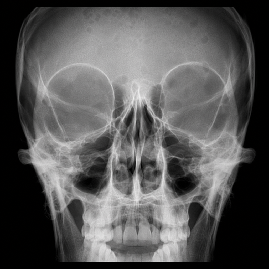
Which of the following eye signs is most likely seen in a patient whose X-ray is shown below?
What condition is characterized by a true pulse in the retinal arteries?
A patient presents with a normal anterior chamber and hazy cornea in one eye, and a shallow anterior chamber and miotic pupil in the fellow eye. Busacca nodules on the iris are observed. What is the diagnosis?
What is the most common etiology of uveitis?
Which of the following signs is characterized by infrequent blinking of the eyelids in Graves' ophthalmopathy?
Which of the following is a feature of Osteogenesis imperfecta?
Practice by Chapter
Diabetes Mellitus
Practice Questions
Hypertension
Practice Questions
Autoimmune Disorders
Practice Questions
Thyroid Disease
Practice Questions
HIV and AIDS
Practice Questions
Hematological Disorders
Practice Questions
Neurological Disorders
Practice Questions
Dermatological Conditions
Practice Questions
Pregnancy-Related Eye Changes
Practice Questions
Metabolic Disorders
Practice Questions
Ocular Toxicity of Systemic Medications
Practice Questions
Infectious Systemic Diseases
Practice Questions
Want unlimited practice?
Get full access to all questions, explanations, and performance tracking.
Scan to download app