Copper deposition in the cornea leads to what finding?
What is Hutchinson's rule in relation to herpes zoster ophthalmicus?
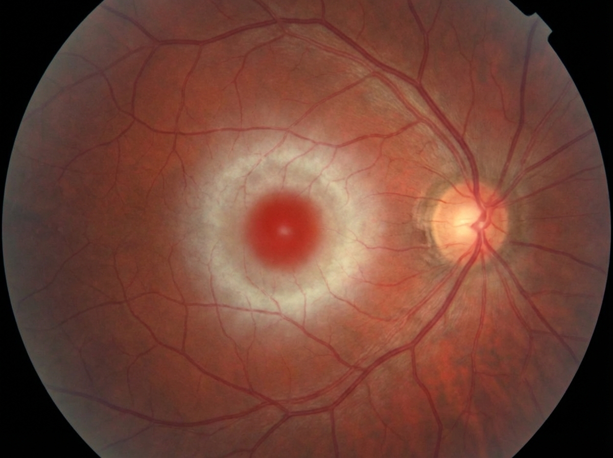
In which of the following diseases is the eye finding shown below seen?

Roth spots are commonly seen in:
What is the most common complication of acute anterior uveitis?
A positive fluorescein dye disappearance test indicates that watering of the eye is due to which of the following?
What is the typical pupil appearance in an acute anterior uveitis attack?
Mutton fat keratic precipitate and Busacca nodules are seen in which type of uveitis?
What is the commonest infection causing blindness in adult men?
A patient presents with freckling in the axilla and inguinal area since childhood. Which of the following is NOT an ophthalmological examination finding in such a patient?
Explanation: **Explanation:** The **Kayser-Fleischer (KF) ring** is the hallmark ocular manifestation of **Wilson’s Disease** (Hepatolenticular degeneration). This condition is characterized by a deficiency in ceruloplasmin, leading to the systemic accumulation of free copper. In the eye, copper is deposited specifically in the **Descemet’s membrane** of the peripheral cornea. It typically appears as a golden-brown or greenish-brown ring, starting superiorly, then inferiorly, and eventually becoming circumferential. It is best visualized using a slit-lamp examination. **Analysis of Incorrect Options:** * **Keratoconus:** A non-inflammatory, progressive thinning and cone-like protrusion of the cornea. While it features a "Fleischer ring" (iron deposition at the base of the cone), it is not caused by copper deposition. * **Keratoglobus:** A rare genetic condition characterized by generalized thinning and hemispherical protrusion of the entire cornea from birth; it is not associated with metallic deposits. * **Siderosis:** This refers to the intraocular deposition of **iron**, usually following a retained iron-containing foreign body. It leads to a rusty discoloration of the iris and lens (Siderosis bulbi), not a copper ring. **High-Yield Clinical Pearls for NEET-PG:** * **Location:** KF rings are found in the **Descemet’s membrane** (not the epithelium or stroma). * **Reversibility:** The ring may disappear with effective chelation therapy (e.g., D-Penicillamine). * **Sunflower Cataract:** Another ocular sign of Wilson’s disease where copper deposits in the **anterior lens capsule**. * **Chalcosis:** The term for tissue reaction to intraocular copper (e.g., from a copper-containing foreign body).
Explanation: ### Explanation **Hutchinson’s Rule** is a clinical sign used to predict the likelihood of ocular involvement in **Herpes Zoster Ophthalmicus (HZO)**. It states that if vesicles appear on the side or tip of the nose, there is a significantly higher risk of intraocular inflammation. **1. Why Option B is Correct:** The rule is based on the anatomy of the **Trigeminal nerve (CN V)**. The Ophthalmic division (V1) branches into the frontal, lacrimal, and **nasociliary nerves**. The nasociliary nerve provides sensory innervation to both the **eyeball** (cornea, iris, and ciliary body) and the **skin of the tip and side of the nose** (via the external nasal nerve). Therefore, cutaneous involvement of the nose indicates that the nasociliary nerve is affected, increasing the probability that the ocular structures are also involved. **2. Why Other Options are Incorrect:** * **Option A:** This is the opposite of the rule. Vesicles on the nose imply a *higher* frequency of ocular involvement, not lower. * **Option C:** While a strong predictor, it is not 100% accurate. Approximately 50–75% of patients with a positive Hutchinson’s sign develop ocular complications, and conversely, the eye can be involved even if the sign is negative. **High-Yield Clinical Pearls for NEET-PG:** * **Causative Agent:** Varicella Zoster Virus (VZV) reactivation in the Gasserian ganglion. * **Most Common Ocular Feature:** Keratitis (specifically **pseudodendrites**, which are stuck-on, peripheral, and lack terminal bulbs, unlike HSV dendrites). * **Treatment:** Oral Acyclovir (800 mg 5 times daily for 7–10 days) started within 72 hours of rash onset. * **Chronic Complication:** Post-herpetic neuralgia is the most common debilitating complication in the elderly.
Explanation: ***GM1 gangliosidosis*** - The **cherry-red spot** seen in the macula is a pathognomonic finding in **GM1 gangliosidosis**, caused by **ganglioside accumulation** in retinal ganglion cells. - This lysosomal storage disorder results in **neurodegeneration** with the fovea appearing red against the surrounding pale retina due to **lipid deposits**. *Gaucher disease* - Typically presents with **pingueculae** (yellowish conjunctival deposits) rather than cherry-red spots. - Ocular manifestations are less common and do not involve the **macular region** with cherry-red appearance. *Von Gierke disease* - This **glycogen storage disease** primarily affects the liver and does not cause **cherry-red spots**. - Ocular findings are not a characteristic feature of this **glucose-6-phosphatase deficiency**. *Galactosemia* - The classic ocular finding is **bilateral cataracts**, particularly **oil drop cataracts** in infants. - Does not cause **cherry-red spots** but rather lens opacification due to **galactitol accumulation**.
Explanation: **Explanation:** **Roth spots** are oval-shaped retinal hemorrhages characterized by a pale, white center. Pathologically, the white center represents a fibrin-platelet thrombus at the site of capillary rupture. **Why Sepsis is the Correct Answer:** While classically associated with **Subacute Bacterial Endocarditis (SBE)**, Roth spots are not pathognomonic for it. They are frequently seen in conditions involving capillary fragility and systemic inflammation, such as **Sepsis**, Leukemia, and severe Anemia. In Sepsis, circulating immune complexes or septic emboli cause localized vasculitis and capillary rupture, leading to the characteristic "white-centered hemorrhage." **Analysis of Incorrect Options:** * **A. Papilledema:** Characterized by optic disc swelling, blurred margins, and splinter (Paton’s) hemorrhages, but not typically Roth spots. * **C. Diabetes:** Diabetic retinopathy presents with microaneurysms, dot-and-blot hemorrhages, and hard exudates. While rare cases exist, it is not the primary association tested. * **D. Hypertension:** Hypertensive retinopathy is marked by arteriolar narrowing, AV nipping, and flame-shaped hemorrhages. **High-Yield Clinical Pearls for NEET-PG:** * **Mnemonic for Roth Spots (LAMES):** **L**eukemia, **A**nemia, **M**ultiple Myeloma/Bacterial Endocarditis, **E**levated Venous Pressure, **S**epsis/Scurvy. * **Differential Diagnosis:** Always look for **Leukemia** if Sepsis or SBE is not in the options, as Leukemia is a very common cause of Roth spots in clinical vignettes. * **Pathology:** The white center is **not** pus; it is a plug of fibrin and platelets (and sometimes leukemic cells).
Explanation: **Explanation:** **Why Cataract is the Correct Answer:** Cataract (specifically **Complicated Cataract**) is the most common complication of recurrent or chronic anterior uveitis. It occurs due to two primary mechanisms: 1. **Inflammatory mediators:** The presence of inflammatory cells and cytokines in the aqueous humor alters the metabolism of the lens fibers. 2. **Steroid-induced:** Long-term use of topical or systemic corticosteroids (the mainstay of treatment) significantly increases the risk of posterior subcapsular cataracts. *Note: Posterior synechiae are a common structural complication, but among the options provided, cataract is the most frequent clinical sequela leading to vision loss.* **Analysis of Incorrect Options:** * **A. Acute congestive glaucoma:** While uveitis can cause "Uveitic Glaucoma" (due to trabeculitis or pupillary block from seclusio pupillae), it is less common than cataract. Acute congestive glaucoma typically refers to primary angle-closure, which is a different entity. * **C. Retinal detachment:** This is a rare complication of anterior uveitis. It is more commonly associated with posterior uveitis or CMV retinitis. * **D. Vitritis:** This refers to inflammation in the vitreous. While "spill-over" cells can be seen in the anterior vitreous in severe anterior uveitis, it is a sign of inflammation rather than a primary long-term complication. **High-Yield Clinical Pearls for NEET-PG:** * **Most common cause of Anterior Uveitis:** Idiopathic (overall); HLA-B27 associated (systemic). * **Cystoid Macular Edema (CME):** The most common cause of **decreased vision** in patients with chronic uveitis. * **Band-shaped Keratopathy:** A classic complication seen specifically in **Juvenile Idiopathic Arthritis (JIA)** associated uveitis. * **Mydriatic of choice:** Atropine 1% is used to provide ciliary rest and prevent/break posterior synechiae.
Explanation: ### Explanation The **Fluorescein Dye Disappearance Test (FDDT)** is a simple clinical test used to evaluate the functional patency of the lacrimal drainage system. **Why Option C is Correct:** The test involves instilling a drop of 2% fluorescein into the conjunctival fornix and observing the tear film for 5 minutes. * **Normal Result:** The dye disappears (drains) within 5 minutes. * **Positive Result:** The dye persists in the conjunctival sac, indicating a **failure of the lacrimal pump mechanism**. This failure occurs in two primary scenarios: 1. **Mechanical Obstruction:** A physical blockage anywhere from the puncta to the nasolacrimal duct prevents the dye from flowing out. 2. **Atony of the Lacrimal Sac:** Even if the passages are physically open, if the sac is atonic (loss of muscle tone/pump action), it cannot "suck" the tears into the drainage system. Therefore, a positive FDDT confirms that the watering is **obstructive/functional** in nature rather than due to hypersecretion (lacrimation). **Why Other Options are Incorrect:** * **Options A & B:** These are incomplete. While both are causes of a positive test, the FDDT cannot differentiate between a physical blockage and a pump failure; it simply indicates that the drainage system is not functioning. * **Option D:** This would describe a negative test result, which is seen in normal eyes or in cases of primary hypersecretion (e.g., ocular surface irritation). **High-Yield Clinical Pearls for NEET-PG:** * **FDDT vs. Jones Test:** FDDT is a non-invasive screening test. If FDDT is positive, the **Jones Dye Test** is performed to localize the site of obstruction. * **Jones Test I:** Differentiates between partial obstruction and hypersecretion. * **Jones Test II (Probing/Irrigation):** Differentiates between upper (canalicular) and lower (nasolacrimal duct) system obstruction. * **Dacryocystography (DCG):** The gold standard for anatomical localization of the site of obstruction.
Explanation: In acute anterior uveitis (iridocyclitis), the pupil is typically **constricted (miotic) and reacts sluggishly** to light. This occurs due to two primary mechanisms: 1. **Ciliary Muscle Spasm:** Inflammation causes irritation and spasm of the ciliary muscle and the sphincter pupillae. 2. **Iris Edema and Engorgement:** The iris becomes swollen and heavy with inflammatory cells and protein-rich fluid (aqueous flare), hindering its mobility. **Explanation of Options:** * **Option D (Correct):** Constriction is a hallmark sign. The "sluggish" reaction is due to the mechanical resistance caused by iris edema and the presence of inflammatory exudates in the anterior chamber. * **Option A & B:** A **semidilated, vertically oval, and fixed** pupil is characteristic of **Acute Congestive Glaucoma**, not uveitis. This is a critical clinical differentiator. * **Option C:** While the pupil can become **irregular** in uveitis, this usually occurs later due to the formation of **posterior synechiae** (adhesions between the iris and the lens). In a fresh, acute attack before synechiae form, the primary finding is simple constriction. **High-Yield Clinical Pearls for NEET-PG:** * **Mydriatics in Uveitis:** Atropine (1%) is the drug of choice. It relieves pain by paralyzing the ciliary muscle (cycloplegia) and prevents the formation of posterior synechiae by keeping the pupil dilated. * **Festooned Pupil:** If posterior synechiae form and a mydriatic is then instilled, the pupil dilates irregularly at non-adherent points, creating a "festooned" appearance. * **Triad of Anterior Uveitis:** Ciliary congestion, Keratic Precipitates (KPs) on the corneal endothelium, and Aqueous Flare/Cells.
Explanation: **Explanation:** The presence of **Mutton-fat Keratic Precipitates (KPs)** and **Busacca nodules** is a hallmark of **Granulomatous Uveitis**. 1. **Why Option A is correct:** Granulomatous inflammation is characterized by a cellular infiltrate of epithelioid cells and macrophages. When these cells settle on the corneal endothelium, they form large, greasy, yellowish-white clusters known as **Mutton-fat KPs**. Furthermore, inflammatory cells can aggregate on the iris surface: **Koeppe nodules** (at the pupillary margin) and **Busacca nodules** (on the iris stroma). These findings are typically seen in systemic conditions like Sarcoidosis, Tuberculosis, Syphilis, and Vogt-Koyanagi-Harada (VKH) syndrome. 2. **Why other options are incorrect:** * **Non-granulomatous uveitis:** Characterized by small, fine, "dust-like" KPs composed mainly of lymphocytes and neutrophils. It lacks the large greasy KPs and iris nodules. * **Posterior uveitis:** This refers to the anatomical location (retina/choroid). While granulomatous diseases can cause posterior uveitis, the specific signs mentioned (KPs and iris nodules) are manifestations of **anterior segment** involvement. * **Choroiditis:** This is a specific form of posterior uveitis involving the choroid; it does not primarily present with corneal precipitates or iris nodules. **High-Yield Clinical Pearls for NEET-PG:** * **Koeppe Nodules:** Found at the pupillary margin (seen in both granulomatous and non-granulomatous). * **Busacca Nodules:** Found on the iris stroma (pathognomonic for **granulomatous** uveitis). * **Arlt’s Triangle:** The typical triangular distribution of KPs on the inferior corneal endothelium due to convection currents in the aqueous humor. * **Total Posterior Synechiae:** Can lead to *Iris Bombe* and secondary angle-closure glaucoma.
Explanation: **Explanation:** **Toxoplasma gondii** is the most common cause of posterior uveitis and infectious retinitis worldwide. In adults, ocular toxoplasmosis typically presents as a reactivation of a congenital infection or a newly acquired infection. It is characterized by a "headlight in the fog" appearance (active retinochoroiditis with overlying vitritis). It remains the leading infectious cause of focal necrotizing retinitis and subsequent permanent visual loss in immunocompetent adults. **Analysis of Incorrect Options:** * **Toxocara (Option A):** Causes Ocular Larva Migrans, typically seen in children who have contact with puppies (T. canis). It usually presents as a unilateral posterior pole granuloma or endophthalmitis, but it is less common than Toxoplasmosis. * **Plasmodium (Option B):** While Malaria is a major systemic infection, its ocular manifestations (retinal hemorrhages, cotton wool spots) are complications of severe/cerebral malaria rather than a primary cause of chronic blindness in the general adult population. * **Taenia solium (Option C):** Causes Cysticercosis. While *Cysticercus cellulosae* can lodge in the subretinal space or vitreous, it is a less frequent cause of blindness compared to the global prevalence of Toxoplasmosis. **NEET-PG High-Yield Pearls:** * **Classic Lesion:** A pigmented "punched-out" chorioretinal scar is the hallmark of old/healed Toxoplasmosis. * **Treatment of Choice:** Triple therapy (Pyrimethamine, Sulfadiazine, and Folinic acid) plus systemic steroids (only after 24-48 hours of antimicrobial cover). * **Congenital Triad (Sabin’s Triad):** Chorioretinitis, Hydrocephalus, and Intracranial calcifications. * **Most common site:** The macula is frequently involved, leading to significant permanent central vision loss.
Explanation: **Explanation:** The clinical presentation of axillary and inguinal freckling (Crowe’s sign) is pathognomonic for **Neurofibromatosis Type 1 (NF1)**, also known as von Recklinghausen disease. NF1 is an autosomal dominant multisystem disorder caused by a mutation in the *NF1* gene on chromosome 17. **Why Aniridia is the Correct Answer:** **Aniridia** (Option D) is the absence of the iris. It is typically associated with mutations in the **PAX6 gene** and is clinically linked to **WAGR syndrome** (Wilms tumor, Aniridia, Genitourinary anomalies, and intellectual disability). It is not a feature of Neurofibromatosis. **Analysis of Incorrect Options (Features of NF1):** * **Optic Nerve Glioma (Option A):** This is a Grade I pilocytic astrocytoma and occurs in approximately 15% of NF1 patients, usually appearing in the first decade of life. * **Lisch Nodules (Option B):** These are melanocytic hamartomas of the iris. They are the most common ocular finding in NF1 and are present in nearly all affected adults. * **Choroidal Nevi/Hamartomas (Option C):** Recent studies and OCT imaging have shown that multifocal choroidal abnormalities (bright patches) are highly sensitive diagnostic markers for NF1. **High-Yield Clinical Pearls for NEET-PG:** * **NF1 (Chromosome 17):** Diagnostic criteria include ≥6 Café-au-lait spots, Lisch nodules, Optic glioma, and sphenoid wing dysplasia. * **NF2 (Chromosome 22):** Characterized by bilateral acoustic neuromas. The classic ocular finding is **PSC (Posterior Subcapsular Cataract)** or combined hamartoma of the retina and RPE. * **Pulsating Proptosis:** In NF1, this occurs due to **sphenoid wing dysplasia**, which allows transmission of CSF pulsations to the orbit.
Diabetes Mellitus
Practice Questions
Hypertension
Practice Questions
Autoimmune Disorders
Practice Questions
Thyroid Disease
Practice Questions
HIV and AIDS
Practice Questions
Hematological Disorders
Practice Questions
Neurological Disorders
Practice Questions
Dermatological Conditions
Practice Questions
Pregnancy-Related Eye Changes
Practice Questions
Metabolic Disorders
Practice Questions
Ocular Toxicity of Systemic Medications
Practice Questions
Infectious Systemic Diseases
Practice Questions
Get full access to all questions, explanations, and performance tracking.
Start For Free