Angioid streaks are seen in which of the following conditions?
Koeppe's nodules are seen in which of the following conditions?
Anterior uveitis is characterized by which of the following?
A chalazion is caused by the obstruction of which gland?
Which of the following is NOT a feature of Behcet's disease?
All of the following can lead to iris neovascularization except?
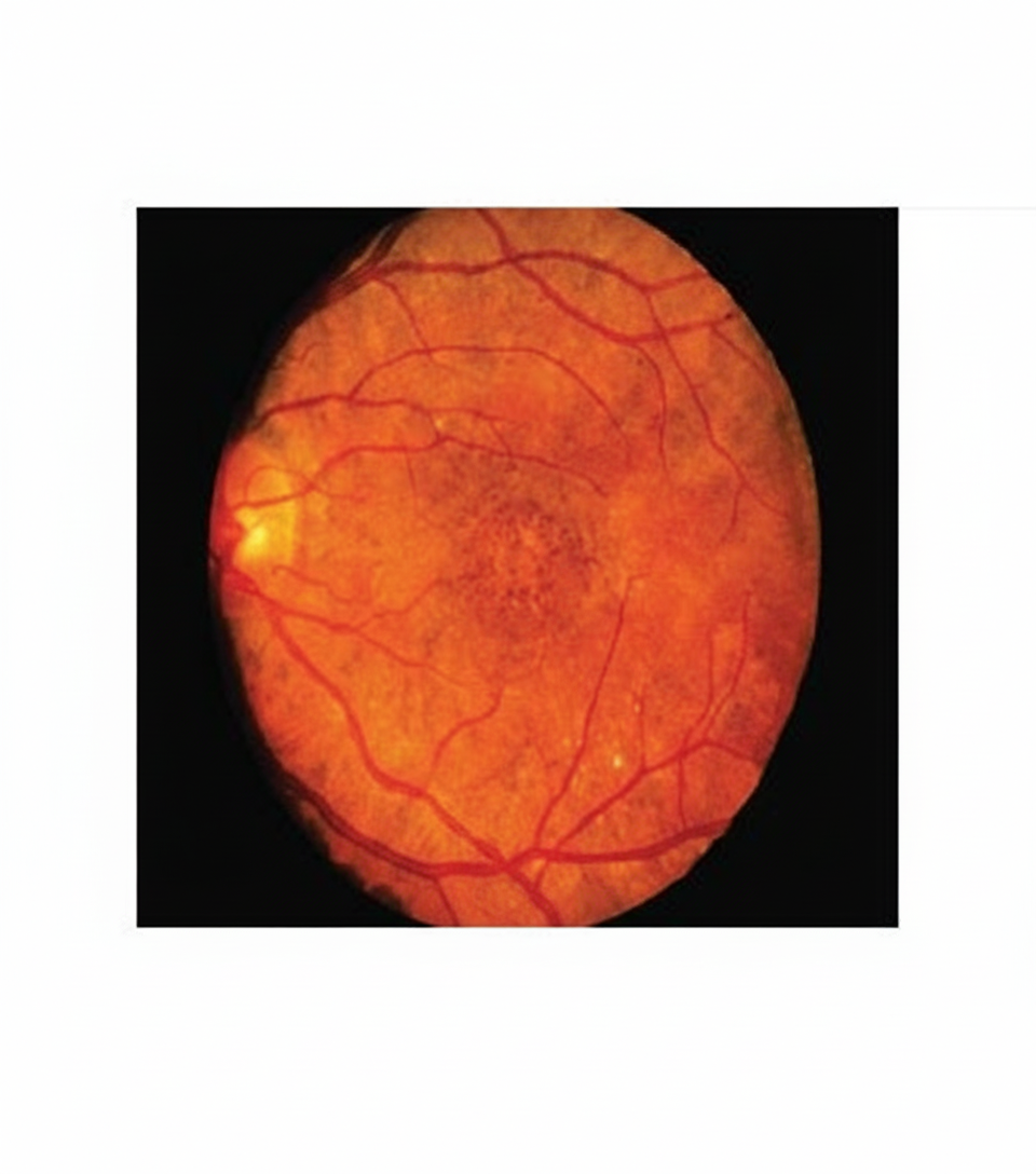
Which of the following metabolic disorders is associated with the fundoscopic finding shown in the image?

A 68-year-old woman with a history of left ventricular congestive heart failure has had decreased visual acuity for the past 5 years. She has no ocular pain and her intraocular pressure is normal. Funduscopic examination reveals arteriolar narrowing, flame-shaped hemorrhages, cotton-wool spots, and hard, waxy exudates. Which of the following underlying diseases is she most likely to have?
What is the most common ocular manifestation in rheumatoid arthritis?
The presence of Kayser-Fleischer rings is pathognomonic of which condition?
Explanation: **Explanation:** Angioid streaks are crack-like dehiscences in a thickened, calcified, and brittle **Bruch’s membrane**. While they appear as irregular, jagged, reddish-brown lines radiating from the peripapillary area, they are often markers of systemic pathology. **Why "All of the above" is the correct clinical context (and why Option D is the intended answer):** The question asks for conditions where angioid streaks are seen. In clinical practice and standard textbooks (like Kanski), angioid streaks are classically associated with the mnemonic **PEPSI**: * **P – Pseudoxanthoma Elasticum (PXE):** The most common association (Grönblad-Strandberg syndrome). * **E – Ehlers-Danlos Syndrome:** Due to connective tissue defects. * **P – Paget’s Disease of bone:** Seen in about 1-2% of cases due to bone metabolism changes. * **S – Sickle Cell Anemia:** And other hemoglobinopathies like Thalassemia. * **I – Idiopathic:** No systemic cause found. **Analysis of Options:** * **A & C (PXE and Paget’s):** These are classic, high-yield causes of angioid streaks. * **B (Acromegaly):** While much rarer than PXE, Acromegaly is a documented systemic association of angioid streaks due to alterations in connective tissue and growth hormone effects. * **D (All of the above):** Since A, B, and C are all recognized associations, "All of the above" is the most comprehensive and correct choice. **NEET-PG High-Yield Pearls:** 1. **Most common cause:** Pseudoxanthoma Elasticum (PXE). 2. **Vision Loss:** The primary cause of vision loss in these patients is **Choroidal Neovascularization (CNV)** occurring through the cracks in Bruch's membrane. 3. **Appearance:** They mimic blood vessels but lie deeper (sub-retinal) and do not follow the normal branching pattern of retinal vasculature. 4. **Mnemonic:** Remember **"PEPSI"** for your exams to quickly recall the systemic associations.
Explanation: **Explanation:** **Koeppe’s nodules** are small, cellular aggregates found on the **pupillary margin** of the iris. They are a hallmark clinical sign of **Granulomatous Uveitis**. 1. **Why Granulomatous Uveitis is correct:** Granulomatous inflammation is characterized by the collection of epithelioid cells, lymphocytes, and multinucleated giant cells. In the eye, these inflammatory cells clump together to form nodules on the iris. Koeppe’s nodules are located at the pupillary border, whereas **Busacca’s nodules** (also seen in granulomatous uveitis) are located on the iris stroma away from the pupil. Common causes include Sarcoidosis, Tuberculosis, and Syphilis. 2. **Why the other options are incorrect:** * **Non-granulomatous uveitis:** This typically presents with a diffuse cellular infiltrate and fine Keratic Precipitates (KPs) rather than organized nodules. * **Choroiditis:** This is a form of posterior uveitis involving the choroid. While it can be granulomatous, iris nodules are specific to anterior segment involvement (iridocyclitis). * **Pars planitis:** This is a subset of intermediate uveitis characterized by "snowbanking" and "snowballs" in the vitreous, not iris nodules. **High-Yield Clinical Pearls for NEET-PG:** * **Koeppe’s nodules:** Pupillary border (Think: **K**oeppe = **K**orner/Edge). * **Busacca’s nodules:** Iris surface/stroma (Pathognomonic for granulomatous disease). * **Mutton-fat Keratic Precipitates (KPs):** Large, greasy-looking inflammatory deposits on the corneal endothelium, also diagnostic of granulomatous uveitis. * **Berlin’s Nodules:** Found in the angle of the anterior chamber (seen on gonioscopy).
Explanation: **Explanation:** The question asks for the feature that is **NOT** characteristic of anterior uveitis (implied by the selection of the "incorrect" clinical sign as the correct answer option). **Why "Shallow Anterior Chamber" is the Correct Answer:** In acute anterior uveitis, the anterior chamber (AC) is typically **normal or deep**. A shallow anterior chamber is a hallmark of **Primary Angle Closure Glaucoma (PACG)**, not uveitis. In fact, in uveitis, the AC may appear deeper than normal due to the breakdown of the blood-aqueous barrier and inflammatory changes. If a shallow AC is seen in a uveitis patient, it usually indicates a complication like *seclusio pupillae* leading to iris bombe, rather than the primary disease process itself. **Analysis of Incorrect Options (Features of Anterior Uveitis):** * **Aqueous Flare:** This is a cardinal sign of anterior uveitis. It is caused by protein leakage from inflamed iris and ciliary body vessels into the aqueous humor (Tyndall effect). * **Circumcorneal Congestion:** Also known as ciliary flush, this is a classic sign of intraocular inflammation. It presents as a purplish-red discoloration around the limbus due to the engorgement of deep ciliary vessels. * **Miosis:** Inflammation causes irritation and spasm of the iris sphincter muscle, leading to a constricted, sluggishly reacting pupil. This also increases the risk of posterior synechiae. **High-Yield Clinical Pearls for NEET-PG:** * **Keratic Precipitates (KPs):** Cellular deposits on the corneal endothelium; "Mutton-fat" KPs suggest granulomatous uveitis (e.g., Sarcoidosis, TB). * **Hypopyon:** A collection of inflammatory cells (pus) in the inferior angle of the AC; commonly seen in HLA-B27 associated uveitis or Behçet’s disease. * **Triad of Iritis:** Pain, photophobia, and blurred vision. * **Differential Diagnosis:** Always differentiate Uveitis (miotic pupil, normal/low IOP) from Acute Congestive Glaucoma (mid-dilated pupil, very high IOP, shallow AC).
Explanation: **Explanation:** A **Chalazion** is a chronic, non-infectious, granulomatous inflammation of the **Meibomian gland**. It occurs due to the obstruction of the gland's duct, leading to the retention of lipid secretions (sebum). This leakage of lipids into the surrounding tarsal plate triggers a sterile "foreign body" inflammatory reaction. **Analysis of Options:** * **A. Meibomian gland (Correct):** These are modified sebaceous glands located within the tarsal plate. Because they are embedded deep in the lid, a chalazion typically presents as a firm, painless nodule pointing towards the conjunctival side. * **B. Zeis's gland:** These are small sebaceous glands associated with the hair follicles of the eyelashes. Infection or inflammation of the Glands of Zeis (or Glands of Moll) results in a **Hordeolum Externum (Stye)**, which is acute, painful, and superficial, unlike a chalazion. * **C. Lacrimal gland:** This gland is responsible for the production of the aqueous layer of the tear film and is located in the superolateral orbit. Inflammation here is termed **Dacryoadenitis**, which presents with lateral upper lid swelling and a characteristic "S-shaped" ptosis. **High-Yield Clinical Pearls for NEET-PG:** * **Pathology:** Histologically, a chalazion shows a **lipogranuloma** (giant cells, epithelioid cells, and lymphocytes surrounding lipid spaces). * **Treatment:** Small ones may resolve spontaneously; larger ones require **Incision and Curettage (I&C)** via a vertical incision (to avoid damaging adjacent Meibomian glands). * **Red Flag:** Recurrent chalazion in the same location in elderly patients should be biopsied to rule out **Sebaceous Gland Carcinoma**.
Explanation: **Explanation:** Behcet’s disease is a chronic, multi-system inflammatory disorder characterized by a relapsing systemic vasculitis. **1. Why "Granulomatous Uveitis" is the correct answer:** Behcet’s disease typically presents with **Non-granulomatous uveitis**. The hallmark ocular finding is a **sterile transient hypopyon** (pus in the anterior chamber that shifts with head position). In contrast, granulomatous uveitis is characterized by large "mutton-fat" keratic precipitates and nodules on the iris, which are typical of conditions like Sarcoidosis or Tuberculosis, not Behcet’s. **2. Analysis of Incorrect Options:** * **Oral Ulcers:** These are the most common and usually the first sign of the disease. They are painful, recurrent, and aphthous-like. * **Erythema Multiforme (Incorrectly listed in options):** While Behcet's is classically associated with **Erythema Nodosum**, some classifications and older texts include various cutaneous vasculitic lesions. However, in the context of this question, the "non-granulomatous" nature of the uveitis is the definitive "False" feature compared to the others. * **HLA B5 Association:** There is a very strong genetic association between Behcet’s disease and **HLA-B5** (specifically the **HLA-B51** subtype), which is a high-yield fact for competitive exams. **Clinical Pearls for NEET-PG:** * **Classic Triad:** Relapsing oral ulcers, genital ulcers, and iridocyclitis (uveitis). * **Pathergy Test:** A unique diagnostic test where a sterile skin papule or pustule forms 24–48 hours after a needle prick. * **Ocular Complication:** It is a leading cause of blindness due to **obliterative retinal vasculitis** (Frosted branch angiitis). * **Treatment:** Systemic steroids and immunosuppressants (like Azathioprine or Cyclosporine) are mainstay therapies.
Explanation: **Explanation:** Iris neovascularization, also known as **Rubeosis Iridis**, is the formation of pathological new blood vessels on the surface of the iris. This process is primarily driven by **ischemia-induced release of Vascular Endothelial Growth Factor (VEGF)** or chronic inflammation. **Why Neurofibromatosis II (NF-II) is the Correct Answer:** NF-II is characterized by bilateral acoustic neuromas, meningiomas, and specific ocular findings like **juvenile posterior subcapsular cataracts** and retinal hamartomas. It does **not** typically cause iris neovascularization. In contrast, **Neurofibromatosis I (NF-I)** is associated with **Lisch nodules** (melanocytic hamartomas), which are distinct from neovascularization. **Analysis of Incorrect Options:** * **Iris Tumors:** Large or malignant tumors (like iris melanoma) can cause chronic inflammation, localized ischemia, or release angiogenic factors, leading to rubeosis iridis. * **Iris Neovascularization:** This option is tautological; the question asks for causes, and the presence of existing neovascularization is the condition itself. (Note: In some exam formats, this may represent a distractor or a typo for a condition like Central Retinal Vein Occlusion). * **Prader-Willi Syndrome:** While rare, systemic associations and secondary ocular complications in syndromic patients can occasionally lead to vascular changes, though it is a less common cause than ischemic retinopathies. **High-Yield Clinical Pearls for NEET-PG:** * **Most Common Cause:** The #1 cause of Rubeosis Iridis is **Diabetic Retinopathy**, followed by **Central Retinal Vein Occlusion (CRVO)** (specifically the ischemic type, often called "100-day glaucoma"). * **Complication:** The primary danger of iris neovascularization is **Neovascular Glaucoma (NVG)**, caused by the fibrovascular membrane contraction closing the drainage angle. * **Management:** Treatment involves **Pan-retinal Photocoagulation (PRP)** to reduce the ischemic drive and Anti-VEGF injections.
Explanation: ***Mitochondrial disorders*** - **Salt-and-pepper pigmentary retinopathy** is a characteristic fundoscopic finding in mitochondrial disorders, particularly **Kearns-Sayre syndrome**. - These disorders affect retinal pigment epithelium function due to **mitochondrial dysfunction**, leading to scattered pigmentary changes throughout the fundus. *Galactosemia* - Primarily causes **oil-droplet cataracts** in the anterior segment, not posterior segment fundoscopic changes. - Fundoscopic examination is typically **normal** unless secondary complications develop from untreated disease. *Albinism* - Characterized by **hypopigmented fundus** with visible choroidal vessels and **foveal hypoplasia**. - Does not produce the salt-and-pepper pigmentary pattern seen in mitochondrial disorders. *GM1 Gangliosidosis* - Classic fundoscopic finding is a **cherry-red spot** in the macula due to ganglioside accumulation. - This presents as a well-defined red central area, distinctly different from diffuse pigmentary retinopathy.
Explanation: **Explanation:** The patient presents with classic features of **Hypertensive Retinopathy**. The clinical findings described—arteriolar narrowing, flame-shaped hemorrhages, cotton-wool spots, and hard exudates—are hallmark signs of chronic high blood pressure affecting the retinal vasculature. 1. **Why Hypertension is correct:** * **Arteriolar narrowing:** A response to vasospasm and hyaline thickening of the vessel walls. * **Flame-shaped hemorrhages:** Occur in the superficial nerve fiber layer (NFL) due to high intravascular pressure. * **Cotton-wool spots:** Represent micro-infarctions of the NFL (ischemia). * **Hard exudates:** Waxy deposits of lipids/proteins resulting from chronic vascular leakage. * **Systemic Link:** The patient’s history of left ventricular heart failure is a strong clinical indicator of long-standing, poorly controlled hypertension. 2. **Why other options are incorrect:** * **Advanced atherosclerosis:** Primarily causes copper/silver wiring and AV nipping (Salus’s/Gunn’s sign) but does not typically cause acute hemorrhages or cotton-wool spots unless associated with hypertension. * **Cerebral edema:** Leads to papilledema (optic disc swelling). While severe hypertension (Grade IV) causes papilledema, the presence of exudates and hemorrhages across the fundus points specifically to retinopathy. * **Diabetes mellitus:** Characterized by **dot-and-blot hemorrhages** (deep retina) and microaneurysms. Flame-shaped hemorrhages are less common in DM compared to HTN. **High-Yield Clinical Pearls for NEET-PG:** * **Keith-Wagener-Barker Classification:** * **Grade I:** Mild generalized arteriolar narrowing. * **Grade II:** Focal narrowing and AV nicking. * **Grade III:** Hemorrhages, exudates, and cotton-wool spots. * **Grade IV:** All of the above + **Papilledema** (Malignant HTN). * **Macular Star:** Formed by hard exudates arranged radially around the fovea, often seen in Grade III/IV HTN. * **Elschnig spots:** Small black spots surrounded by yellow halos, representing choroidal infarcts in severe hypertension.
Explanation: **Explanation:** **Keratoconjunctivitis sicca (KCS)**, or secondary Sjögren’s syndrome, is the **most common** ocular manifestation of Rheumatoid Arthritis (RA), occurring in approximately 15–25% of patients. It results from immune-mediated destruction of the lacrimal glands, leading to aqueous tear deficiency. Patients typically present with a foreign body sensation, burning, and dryness. **Analysis of Options:** * **B. Episcleritis:** This is the most common **inflammatory** complication of the sclera/episclera in RA, but its overall prevalence is lower than KCS. It is usually benign and self-limiting. * **A. Scleritis:** While RA is the most common systemic cause of scleritis (especially necrotizing scleritis), it is less frequent than both KCS and episcleritis. Its presence often indicates high systemic disease activity and a poor prognosis. * **D. Anterior Uveitis:** This is more characteristically associated with Seronegative Spondyloarthropathies (like Ankylosing Spondylitis) rather than Rheumatoid Arthritis. **High-Yield Clinical Pearls for NEET-PG:** * **Most common ocular finding:** Keratoconjunctivitis sicca. * **Most specific/serious ocular finding:** Necrotizing scleritis (Scleromalacia perforans), which occurs in long-standing RA and is characterized by painless thinning of the sclera without inflammation. * **Drug-induced toxicity:** Patients on Hydroxychloroquine (HCQ) for RA require baseline and periodic screening for **"Bull’s eye maculopathy."** * **Peripheral Ulcerative Keratitis (PUK):** A serious "melting" of the cornea that can occur in severe RA.
Explanation: **Explanation:** **Wilson’s Disease (Hepatolenticular Degeneration)** is the correct answer. The **Kayser-Fleischer (KF) ring** is a pathognomonic sign caused by the deposition of **copper** in the **Descemet’s membrane** of the cornea. It typically appears as a golden-brown or greenish-brown ring at the periphery of the cornea, starting superiorly, then inferiorly, and eventually becoming circumferential. It is seen in 95% of patients with neurological involvement and about 50-60% of those with hepatic involvement. **Analysis of Incorrect Options:** * **Keratoconus:** Characterized by **Fleischer rings** (iron deposition at the base of the cone) and **Munson’s sign**, but not KF rings. * **Lowe’s Syndrome (Oculo-cerebro-renal syndrome):** A rare X-linked recessive disorder characterized by congenital cataracts, glaucoma, and renal tubular dysfunction. It does not feature copper deposition. * **Albinism:** Associated with iris transillumination defects, foveal hypoplasia, and nystagmus due to lack of melanin, but no corneal rings. **High-Yield Clinical Pearls for NEET-PG:** * **Location:** Copper deposits specifically in the **Descemet’s membrane** (not the epithelium or stroma). * **Reversibility:** The KF ring may disappear with effective chelation therapy (e.g., Penicillamine). * **Sunflower Cataract:** Another ocular feature of Wilson’s disease, where copper deposits in the anterior lens capsule. * **Diagnosis:** Best visualized using a **Slit-lamp examination** in early stages. * **Differential:** Do not confuse *Fleischer rings* (Iron in Keratoconus) with *Kayser-Fleischer rings* (Copper in Wilson's).
Diabetes Mellitus
Practice Questions
Hypertension
Practice Questions
Autoimmune Disorders
Practice Questions
Thyroid Disease
Practice Questions
HIV and AIDS
Practice Questions
Hematological Disorders
Practice Questions
Neurological Disorders
Practice Questions
Dermatological Conditions
Practice Questions
Pregnancy-Related Eye Changes
Practice Questions
Metabolic Disorders
Practice Questions
Ocular Toxicity of Systemic Medications
Practice Questions
Infectious Systemic Diseases
Practice Questions
Get full access to all questions, explanations, and performance tracking.
Start For Free