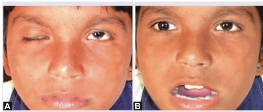
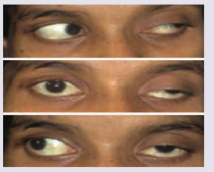
A 10-year-old girl child presents with ptosis of right eyelid. On movement of jaw there is retraction of ptotic eyelid. The image of the patient is shown below. This condition is known as:

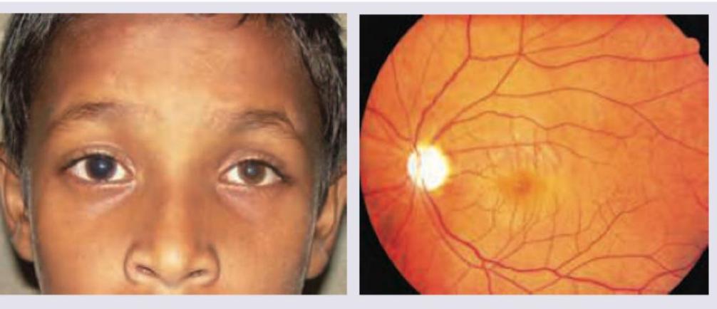
An 8-year-old child presents with gradual reduction in vision in the right eye. Family history of similar presentation was elicited on the maternal side. On examination, on right side direct light reflex is absent and consensual light reflex is present. Fundus examination was performed. What is the diagnosis?

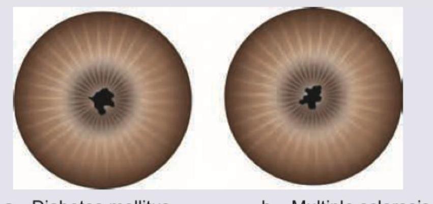
The pupils shown below are miotic and irregular in shape. They are nonreactive to light but react to accommodation. They also do not dilate with atropine. All are causes of these pupils except:

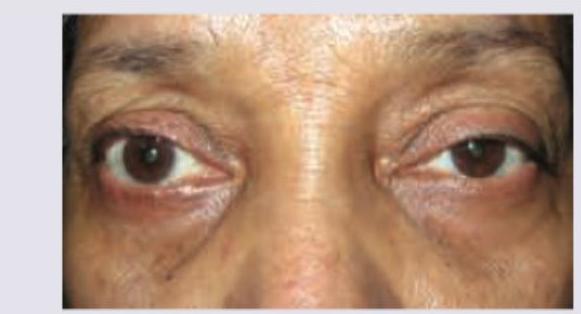
A 25-year-old lady presents with reduced color vision and paresthesias. Her symptoms increase with rise of body temperature. Examine the appearance of her eyes and comment on diagnosis.

The following visual loss is seen due to lesion in:

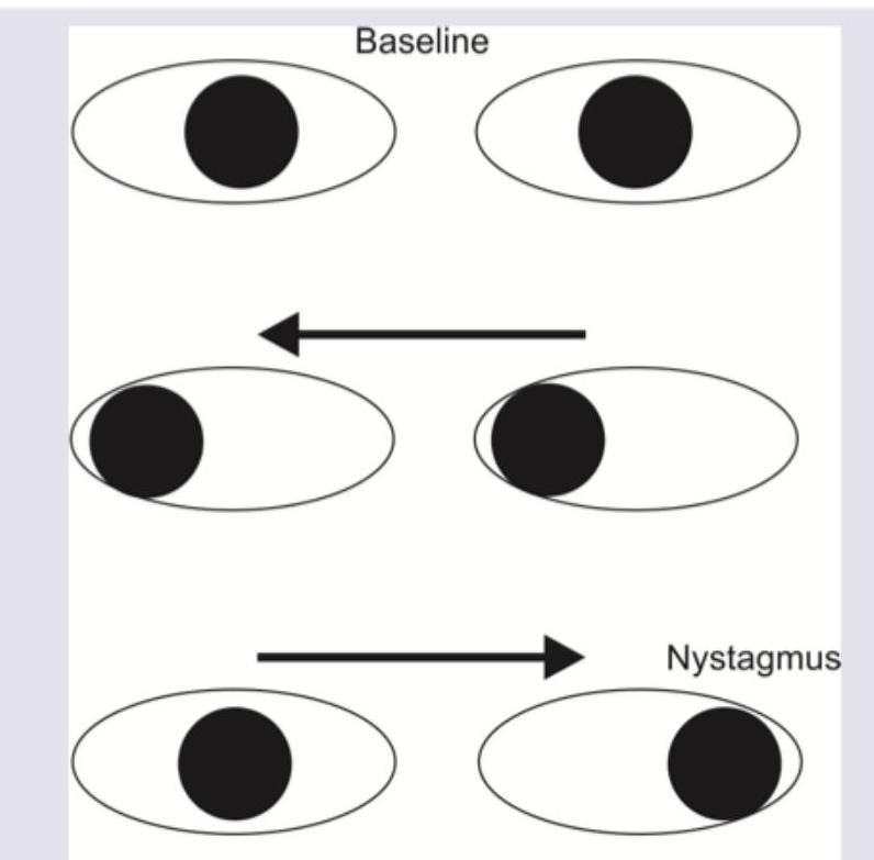
A patient presents with the eye movement abnormalities shown in the image below. What is the diagnosis?

What is the diagnosis?

The given image shows which of the following conditions? (AIIMS Nov 2018)

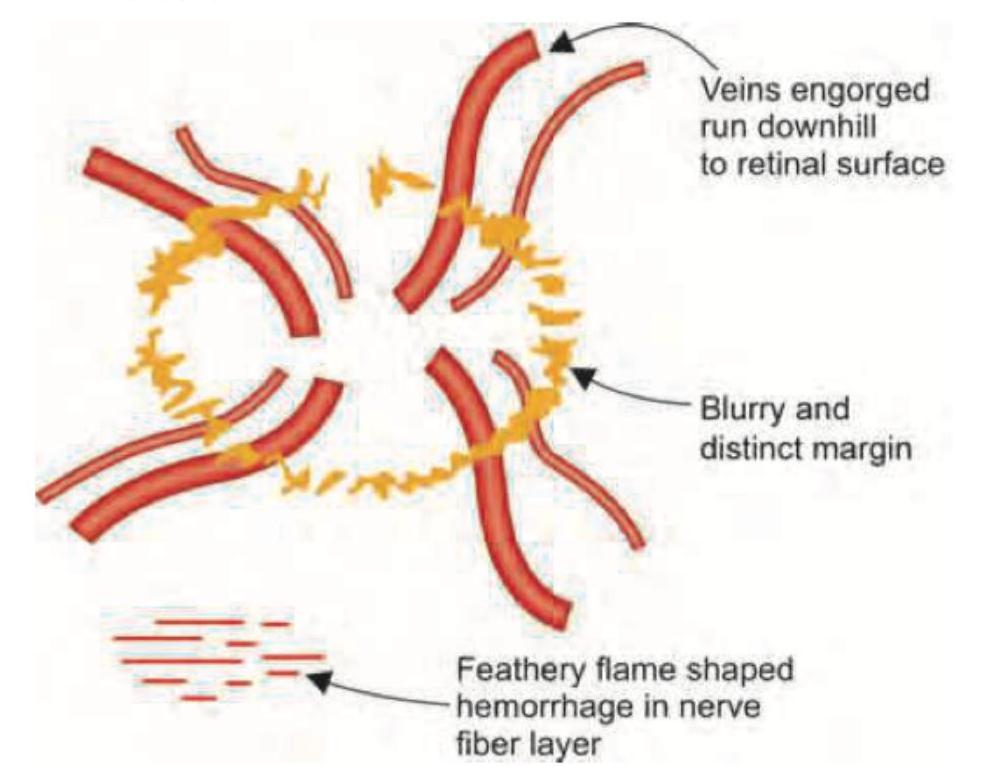
The images show a normal optic disc (left) and papilledema with blurred disc margins (right). What is the most common cause of this condition?

Identify the visual field defect shown in the image.

Explanation: ***Marcus Gunn syndrome*** - This syndrome, also known as the **Marcus Gunn jaw-winking phenomenon**, is characterized by a unilateral congenital ptosis that retracts or elevates when the jaw is moved (e.g., chewing, sucking, jaw protrusion). - The image clearly shows **ptosis of the right eyelid** (Image A) and **retraction of the ptotic eyelid** when the mouth is open (jaw movement, Image B), which is the hallmark of this condition. *Marcus Gunn pupil* - This refers to an **afferent pupillary defect (APD)**, typically detected by the swinging flashlight test, where the pupil paradoxically dilates when light is shined into the affected eye. - It is a sign of **optic nerve damage** and is unrelated to eyelid ptosis or jaw movements affecting the eyelid. *Floppy eyelid syndrome* - This condition is characterized by **loose, elastic upper eyelids** that evert easily, often during sleep, and are associated with chronic papillary conjunctivitis. - It is commonly seen in **obese, middle-aged men** and does not involve jaw-winking phenomena or congenital ptosis. *Myasthenia gravis* - This is an **autoimmune neuromuscular disorder** causing fluctuating weakness of voluntary muscles, including the extraocular muscles and those responsible for eyelid elevation. - While it can cause **ptosis** and **diplopia**, the ptosis typically worsens with fatigue and does not exhibit the specific jaw-winking phenomenon.
Explanation: ***Optic atrophy*** - The clinical finding of **gradual reduction in vision**, **absent direct light reflex** (indicating an afferent pupillary defect), and **present consensual light reflex** in the affected eye confirms optic nerve pathology. - The fundus image shows **optic disc pallor**, indicating loss of retinal ganglion cell axons, which is characteristic of optic atrophy. - The **positive family history on the maternal side** in an 8-year-old child strongly suggests **hereditary optic atrophy** (such as Leber's Hereditary Optic Neuropathy or Dominant Optic Atrophy), making this the most likely diagnosis. *ICSOL* - **Intracranial space-occupying lesions** can cause compressive optic neuropathy and secondary optic atrophy, but typically present with other neurological signs such as headache, papilledema, or focal neurological deficits. - While possible, the strong family history and isolated unilateral presentation in a child make hereditary optic atrophy more likely than an acquired ICSOL. *Devic's disease* - **Devic's disease** (Neuromyelitis Optica Spectrum Disorder) involves optic neuritis and transverse myelitis, typically presenting with acute, painful, often bilateral vision loss along with spinal cord symptoms. - The gradual, unilateral vision loss with established optic disc pallor suggests chronic nerve damage rather than the acute inflammatory process seen in Devic's disease. - Family history is not a typical feature of NMO. *Iritis* - **Iritis** (anterior uveitis) is an inflammatory condition of the iris characterized by eye pain, redness, photophobia, and decreased vision due to inflammation. - Examination would reveal inflammatory cells in the anterior chamber, circumcorneal congestion, and possibly posterior synechiae—not optic disc pallor. - Iritis does not cause afferent pupillary defects or optic nerve damage as the primary pathology.
Explanation: ***Multiple sclerosis*** - Multiple sclerosis does not directly cause **miotic, irregular, nonreactive to light but reactive to accommodation pupils**, a constellation of signs known as **Argyll Robertson pupils**. While MS can cause various ocular motor disorders and pupillary abnormalities (e.g., afferent pupillary defect from optic neuritis), it typically does not present with this specific pupillary response. - Ocular involvement in **multiple sclerosis** usually includes optic neuritis, nystagmus, and internuclear ophthalmoplegia, which are distinct from the described pupillary findings. - **MS is the least associated** with this classic pupillary presentation among the options listed. *Diabetes mellitus* - **Diabetic autonomic neuropathy** can affect the parasympathetic innervation to the iris sphincter and ciliary body, potentially resulting in **Argyll Robertson-like pupils** that are miotic, irregular, and show light-near dissociation. - While less common than neurosyphilis (the classic cause), diabetes is a recognized cause of **autonomic neuropathy** affecting pupillary reflexes and can produce these findings. *Pinealoma* - A **pinealoma** can cause **Parinaud's syndrome** (dorsal midbrain syndrome), which includes **light-near dissociation** where pupils react to accommodation but not to light. - However, it should be noted that pupils in Parinaud's syndrome are typically **mid-dilated rather than miotic**, making this a less typical cause of the complete constellation described. - Despite this difference, pinealomas are classically taught as a cause of light-near dissociation, which is a key feature of these pupils. *Iridocyclitis* - **Iridocyclitis** (anterior uveitis) causes inflammation of the iris and ciliary body, leading to **posterior synechiae**, where the iris adheres to the lens. - These adhesions cause the pupil to become **irregular in shape and miotic** (due to sphincter spasm and scarring). - The inflamed iris shows diminished reaction to light, and **atropine resistance** occurs due to adhesions preventing pupillary dilation, matching all the described features.
Explanation: ***Internuclear ophthalmoplegia*** - The clinical presentation of **reduced color vision**, **paresthesias**, and symptoms worsening with increased body temperature (**Uhthoff's phenomenon**) is highly suggestive of **multiple sclerosis**. - **Internuclear ophthalmoplegia (INO)** is a common and characteristic ocular manifestation of multiple sclerosis, causing impaired adduction of one eye during conjugate gaze with nystagmus of the abducting eye. Examination of the image would likely show such a gaze abnormality. *Horner syndrome* - **Horner syndrome** is characterized by **miosis**, **ptosis**, and **anhidrosis** on one side of the face. - These specific ocular findings and systemic symptoms (reduced color vision, paresthesias, Uhthoff's phenomenon) do not align with a classic Horner syndrome presentation. *Duane retraction syndrome* - **Duane retraction syndrome** is a congenital disorder characterized by **limited abduction** (most common) or adduction, globe retraction, and narrowing of the palpebral fissure on attempted adduction. - This is a developmental anomaly of ocular motility and does not explain the widespread neurological symptoms or the heat sensitivity described. *Hutchinson pupil* - A **Hutchinson pupil** is a fixed, dilated pupil seen in cases of **uncal herniation** due to compression of the oculomotor nerve (CN III). - This is an acute, unilateral neurological emergency and does not present with the chronic, relapsing-remitting symptoms like reduced color vision and paresthesias or Uhthoff's phenomenon.
Explanation: ***Optic chiasma*** - The image shows **bitemporal hemianopsia**, characterized by loss of vision in the **temporal (outer) halves of both visual fields**. - This specific visual field defect occurs due to damage to the **crossing nasal fibers** within the optic chiasma, which carry information from the temporal visual fields. *Optic tract* - A lesion in the optic tract would cause a **homonymous hemianopsia**, meaning loss of vision in the **same half of the visual field** for both eyes (e.g., right homonymous hemianopsia). - This is because the optic tract contains fibers from the contralateral nasal retina and ipsilateral temporal retina. *Optic radiation* - Damage to the optic radiation typically results in **homonymous quadrantanopia** or **homonymous hemianopsia**, depending on the extent and location of the lesion. - For instance, a lesion in the temporal lobe (Meyer's loop) affects the superior visual field and causes **superior homonymous quadrantanopia**. *Posterior calcarine fissure* - A lesion in the posterior calcarine fissure (primary visual cortex) would cause a **contralateral homonymous hemianopsia with macular sparing**. - Macular sparing often occurs because the macula's representation is usually spared due to its dual blood supply or extensive representation.
Explanation: ***Internuclear ophthalmoplegia*** - The image shows **impaired adduction** of one eye with **abducting nystagmus** in the contralateral eye during horizontal gaze, which is the hallmark of **internuclear ophthalmoplegia (INO)**. - This condition results from damage to the **medial longitudinal fasciculus (MLF)**, which connects the abducens nucleus to the contralateral oculomotor nucleus, disrupting coordinated horizontal conjugate gaze. - In INO, the affected eye fails to adduct past midline, while the abducting eye shows nystagmus. - **Bilateral INO** (as shown) is commonly seen in **multiple sclerosis** in young patients and **brainstem stroke** in older patients. *Left 3rd nerve palsy* - A 3rd nerve palsy would cause **ptosis**, **dilated pupil**, and inability to move the eye in multiple directions (impaired adduction, elevation, and depression). - The pattern shown is specifically an **adduction deficit during conjugate gaze** with preserved convergence (typical of INO), not the complete ophthalmoplegia seen in 3rd nerve palsy. - Abducting nystagmus in the contralateral eye is characteristic of INO, not 3rd nerve palsy. *Horizontal gaze palsy* - A horizontal gaze palsy involves inability of **both eyes** to move conjugately in one horizontal direction due to damage to the **pontine paramedian reticular formation (PPRF)** or **abducens nucleus**. - In the image, one eye can abduct (showing the PPRF and abducens nucleus are intact), while the other eye fails to adduct—this dissociation is characteristic of **INO**, not gaze palsy. *Duane retraction syndrome* - Duane syndrome is a **congenital** disorder with **limited abduction** or adduction, **globe retraction** on attempted adduction, and narrowing of the palpebral fissure. - The key finding of **abducting nystagmus** in the contralateral eye and the pattern of acquired adduction failure are not consistent with Duane syndrome. - Duane syndrome typically presents in childhood, not acutely as shown here.
Explanation: ***Third nerve palsy*** - The image shows **ptosis** (drooping of the eyelid) and **pupil dilation** with the eye deviated **down and out**, which are classic signs of a complete third nerve palsy. - The **oculomotor nerve (CN III)** innervates most extraocular muscles (**superior, medial, inferior recti, and inferior oblique**), the **levator palpebrae superioris**, and the **parasympathetic fibers to the pupillary constrictor muscle**. *Sixth nerve palsy* - A sixth nerve palsy (**abducens nerve**) would cause an inability to **abduct the eye** (move it outwards) leading to an **esotropia** (eye turned inward at rest) and **horizontal diplopia**, which is not depicted. - Symptoms primarily affect the **lateral rectus muscle**. *Myasthenia gravis* - Myasthenia gravis can cause **ptosis** and **diplopia**, but typically presents with **variable and fluctuating weakness** that worsens with fatigue. - It does not usually cause the fixed, specific pattern of strabismus and pupillary involvement seen in a severe third nerve palsy. *Tolosa-Hunt syndrome* - Tolosa-Hunt syndrome is a **painful ophthalmoplegia** caused by **non-specific inflammation** in the cavernous sinus or orbital apex. - While it can affect cranial nerves III, IV, V1, and VI, leading to **ophthalmoplegia** and **ptosis**, the defining feature is often **severe orbital pain**, which cannot be assessed from the image alone, and the specific constellation of symptoms in the image points more directly to a third nerve lesion rather than an inflammatory syndrome.
Explanation: ***Inter-nuclear ophthalmoplegia*** - The image shows that the **right eye** is **unable to adduct past the midline** when attempting to look left (middle image), which is a classic sign of internuclear ophthalmoplegia (INO). - The **left eye** shows **abducting nystagmus**, a common associated finding in INO due to damage to the **medial longitudinal fasciculus (MLF)**. *Oculomotor nerve palsy* - An oculomotor nerve (CN III) palsy would typically present with a **down-and-out deviation** of the affected eye, **ptosis**, and a **dilated pupil**, which are not seen here. - The patient would also have difficulty moving the eye medially, superiorly, and inferiorly, whereas here the right eye can move down and up, but not fully medially. *Lateral rectus palsy* - A lateral rectus (CN VI) palsy would cause an inability to **abduct** the affected eye and an **esotropia** (eye turning inward) at primary gaze. - The image shows impaired adduction, not abduction, of the right eye, ruling out lateral rectus palsy. *Trochlear nerve palsy* - A trochlear nerve (CN IV) palsy results in weakness of the **superior oblique muscle**, leading to **vertical diplopia**, especially when looking down and in, and a characteristic **head tilt** to compensate. - This presentation does not match the image, which primarily demonstrates a horizontal gaze abnormality with adduction deficit.
Explanation: ***ICSOL (Intracranial Space-Occupying Lesion)*** - The image on the right depicts **papilledema**, characterized by **blurred optic disc margins**, elevated disc, venous engorgement, and flame-shaped hemorrhages - Papilledema is a classic sign of **raised intracranial pressure** - **Intracranial space-occupying lesions (ICSOL)** such as brain tumors, abscesses, or hematomas are among the most common causes of increased ICP leading to papilledema - The increased pressure is transmitted to the optic nerve sheath, causing optic disc swelling *Malignant Hypertension* - Can cause hypertensive retinopathy with disc swelling, but typically presents with other systemic features (severely elevated BP >180/120 mmHg) - Would show additional retinal changes like cotton-wool spots, exudates, and arteriolar narrowing *Idiopathic Intracranial Hypertension (Pseudotumor Cerebri)* - Causes raised ICP without a mass lesion - More common in obese women of childbearing age - Important differential but ICSOL is more commonly tested as the classic cause *Central Retinal Vein Occlusion (CRVO)* - Causes disc swelling but presents with extensive retinal hemorrhages in all quadrants ("blood and thunder" appearance) - Due to venous obstruction, not raised ICP - Distinct clinical picture from papilledema
Explanation: ***Bitemporal hemianopia*** - The image shows loss of vision in the **temporal (outer) halves of both visual fields**, which is characteristic of bitemporal hemianopia. - This defect typically results from a lesion at the **optic chiasm**, compressing the crossing nasal retinal fibers, often due to a **pituitary tumor**. *Binasal hemianopia* - This condition involves visual loss in the **nasal (inner) halves of both visual fields**, which is the opposite of what is depicted. - It is a rare defect that can be caused by lesions affecting the **uncrossed temporal retinal fibers** on both sides, such as from bilateral carotid artery aneurysms. *Homonymous hemianopia* - A homonymous hemianopia involves the **same half of the visual field in both eyes** (e.g., right visual field loss in both eyes), resulting from a lesion posterior to the optic chiasm. - The image clearly shows different halves affected in each eye (temporal fields), not the same half. *Altitudinal defect* - An altitudinal defect involves the **loss of vision in the upper or lower half of the visual field** in one or both eyes, respecting the horizontal midline. - The visual field loss shown in the image is vertical, affecting the temporal halves, not the upper or lower halves.
Anatomy of Visual Pathways
Practice Questions
Pupillary Disorders
Practice Questions
Optic Neuritis
Practice Questions
Ischemic Optic Neuropathies
Practice Questions
Other Optic Neuropathies
Practice Questions
Papilledema
Practice Questions
Cranial Nerve Palsies
Practice Questions
Nystagmus
Practice Questions
Visual Field Defects
Practice Questions
Neuro-ophthalmic Manifestations of Intracranial Lesions
Practice Questions
Functional Visual Disorders
Practice Questions
Migraine and the Eye
Practice Questions
Get full access to all questions, explanations, and performance tracking.
Start For Free