A patient with pituitary adenoma compressing the optic chiasma now presents with loss of visual field. What visual field defect will be seen in the patient?
A patient presents with right-sided field defects in both eyes, but central vision remains unaffected. What is the most likely diagnosis?
Which of the following is NOT a feature of Horner's Syndrome?
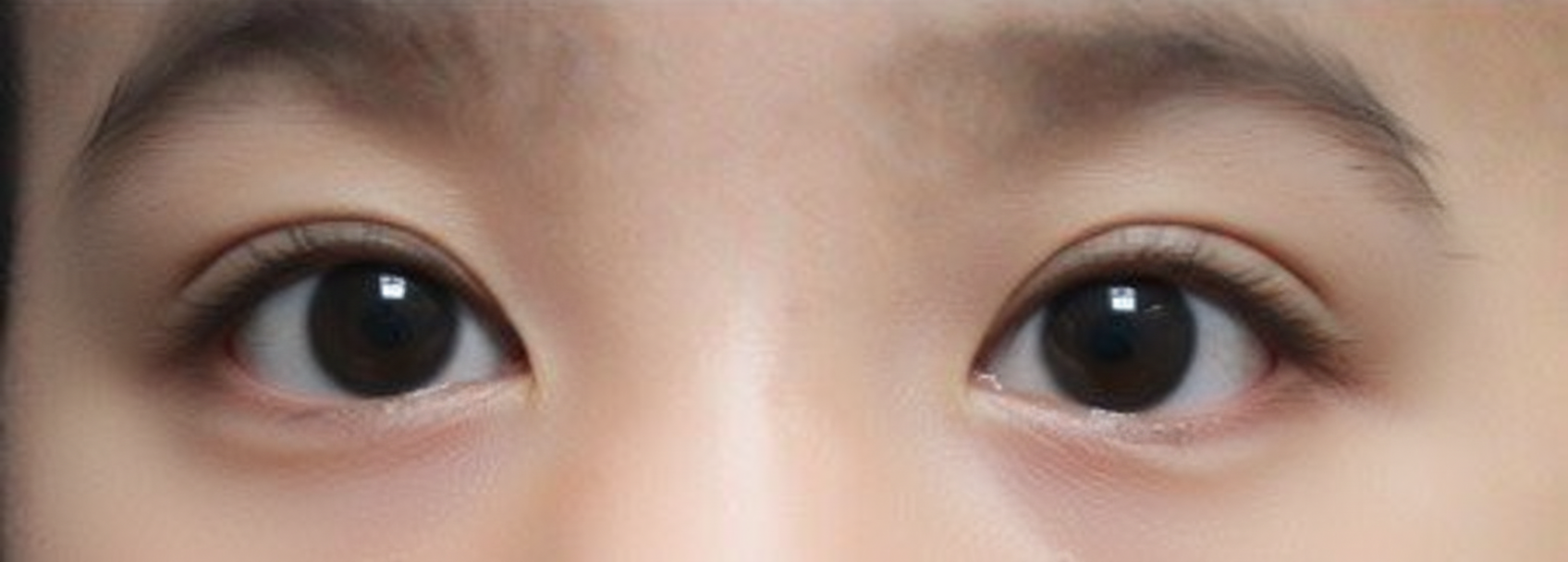
Which of the following is the diagnosis based on the given eye movement abnormality image?
In optic neuritis, which is true?
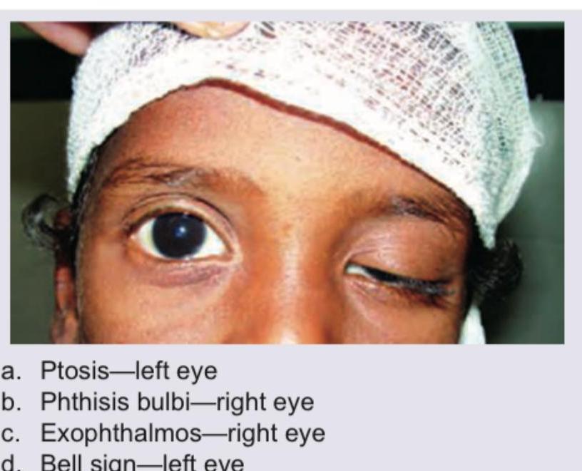
What is the clinical condition shown in the image?

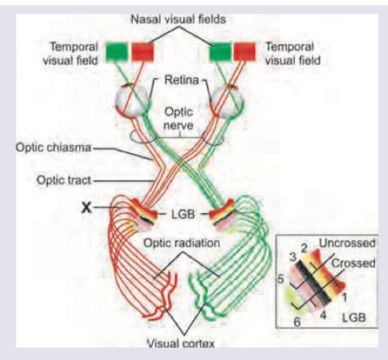
Which of the following lesions will develop after lesion in the area marked as $X$ ?

A child has been diagnosed with cavernous sinus meningioma. Which of these is correct?

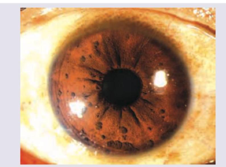
The following image shows:

All are causes of the field defect shown below except:

Explanation: ***Bitemporal hemianopia*** - A **pituitary adenoma** most commonly compresses the **optic chiasma**, where the nasal retinal fibers from both eyes decussate (cross over). - Damage to these crossing fibers specifically leads to the loss of the **temporal visual fields** in both eyes, which is the definition of bitemporal hemianopia. *Homonymous anopia* - This is a less specific term. **Homonymous** defects refer to the loss of the same side of the visual field in both eyes (e.g., the left side in both eyes). - These types of defects are caused by lesions located **posterior** (behind) the optic chiasma, not at the chiasma itself. *Homonymous hemianopia* - This defect describes the loss of the same half (either left or right) of the visual field in both eyes. - It results from a lesion in the visual pathway **posterior** to the optic chiasma, such as in the **optic tract**, **optic radiations**, or the **visual cortex**. *Superior quadrantanopia* - This refers to the loss of vision in the upper quadrant of the visual field, often described as a "pie in the sky" defect. - This is typically caused by a lesion affecting the contralateral **temporal lobe** (damaging **Meyer's loop** of the optic radiation), not the optic chiasma.
Explanation: ***Homonymous Hemianopia with Macular Sparing*** - **Homonymous hemianopia** refers to a visual field defect on the same side in both eyes (in this case, the right side), which precisely matches the patient's presentation. - **Macular sparing** occurs because the occipital pole, which processes central vision, often has a dual blood supply from both the **posterior cerebral artery (PCA)** and the **middle cerebral artery (MCA)**, protecting it during a stroke affecting one vessel. *Heteronymous Hemianopia with Central Sparing* - **Heteronymous hemianopia** involves defects on opposite sides of the visual field in each eye (e.g., loss of both temporal fields), which is inconsistent with the patient's right-sided defect in both eyes. - This type of defect is classically caused by a lesion at the **optic chiasm**, such as a pituitary adenoma, leading to **bitemporal hemianopia**. *Optic Chiasm Lesion* - A lesion compressing the optic chiasm, where the nasal retinal fibers cross, typically causes **bitemporal hemianopia** (loss of peripheral vision in both eyes). - This results in a **heteronymous** defect, not a **homonymous** one as described in the question. *Optic Tract Lesion* - A lesion in the optic tract (posterior to the chiasm) does cause a contralateral **homonymous hemianopia**. - However, lesions in the optic tract typically do not spare the macula, as the fibers from the macula are intermingled with other fibers at this point. Macular sparing points towards a more posterior lesion in the **visual cortex**.
Explanation: ***Exophthalmos*** - **Exophthalmos** (bulging of the eyeball) is caused by hyperactivity of the sympathetic system (e.g., in *hyperthyroidism*) or orbital mass effects, making it *NOT* a feature of sympathetic paralysis. - Horner's Syndrome, due to paralysis of the **Müller's muscle** in the orbit, actually causes the opposite effect: apparent **enophthalmos** (sinking of the eyeball). *Anhidrosis* - **Anhidrosis** (lack of sweating) on the ipsilateral face and neck is a crucial component of Horner's syndrome, particularly if the lesion is located preganglionic or centrally. - This occurs because the sympathetic fibers supplying the **sweat glands** are disrupted along the pathway. *Miosis* - **Miosis** (constricted pupil) is a hallmark feature resulting from the unopposed action of the parasympathetic system's **sphincter pupillae muscle**. - The sympathetic nerves responsible for innervating the **dilator pupillae muscle** are paralyzed, leading to relative pupil constriction that is more pronounced in dim light (*anisocoria*). *Ptosis* - **Ptosis** (droopy eyelid) is a characteristic symptom caused by the paralysis of the sympathetically innervated **Müller's muscle** (superior tarsal muscle). - This results in a mild degree of eyelid drooping (partial ptosis), often less severe than the ptosis seen with **Oculomotor Nerve (CN III) palsy**.
Explanation: **Internuclear ophthalmoplegia** - This diagnosis is indicated by the failure of the right eye to **adduct** (move inwards) when looking to the left, which is a hallmark sign. This specific defect is caused by a lesion in the **Medial Longitudinal Fasciculus (MLF)** on the same side as the adduction failure. - Another key feature shown is **nystagmus** in the contralateral (left) eye during **abduction** (outward movement). This combination of ipsilateral adduction failure and contralateral abducting nystagmus is classic for INO. *3rd nerve palsy* - A 3rd nerve palsy would present with the affected eye positioned 'down and out' due to unopposed action of the superior oblique and lateral rectus muscles. It also typically involves **ptosis** and a **dilated pupil**. - In the given image, the vertical movements and pupillary function are not depicted as abnormal, and the primary issue is with horizontal conjugate gaze, not the multiple deficits seen in 3rd nerve palsy. *6th nerve palsy* - This condition results in the inability to **abduct** the eye (move it outwards) due to paralysis of the **lateral rectus muscle**. The patient would complain of horizontal diplopia, worse on gaze towards the affected side. - The image shows that both eyes are capable of abduction. The defect is clearly in adduction of the right eye. *Horizontal gaze palsy* - This involves the inability of **both eyes** to move in one horizontal direction. It is caused by a lesion in the pontine gaze center, the **Paramedian Pontine Reticular Formation (PPRF)**. - In this case, the left eye successfully moves to the left, and both eyes can move to the right, ruling out a complete gaze palsy to either side.
Explanation: ***Unilateral vision loss, pain on eye movement, and decreased color vision*** - This combination represents the **classic triad** of optic neuritis: sudden **unilateral vision loss**, **pain with eye movement** (present in >90% of cases), and **dyschromatopsia** (decreased color discrimination, especially red-green). - These three features together provide the most comprehensive and accurate clinical picture of typical **inflammatory optic neuritis**, often associated with **multiple sclerosis**. *Unilateral vision loss with decreased color vision* - While **unilateral presentation** and **dyschromatopsia** are indeed characteristic features of optic neuritis, this option omits the highly specific symptom of **pain on eye movement**. - **Pain with eye movement** is present in over 90% of optic neuritis cases and is a key differentiating feature from other causes of **acute vision loss**. *Pain on eye movement only* - Although **retrobulbar pain** with eye movement is highly characteristic and present in most cases, focusing solely on this symptom ignores the primary visual manifestations. - Optic neuritis by definition involves **optic nerve inflammation** causing **visual dysfunction**, including decreased acuity and **color vision deficits**. *Bilateral vision loss is common* - **Bilateral optic neuritis** is actually uncommon in typical cases and suggests **atypical forms** such as **Neuromyelitis Optica Spectrum Disorder (NMOSD)** or **autoimmune optic neuropathy**. - Classic optic neuritis associated with **multiple sclerosis** typically presents **unilaterally**, with the fellow eye remaining unaffected initially.
Explanation: ***Correct: Unilateral ptosis (Right eye)*** - The image clearly shows **drooping of the right upper eyelid**, which is the hallmark of **ptosis** - This is **unilateral**, affecting only the right eye, with the left eye appearing normal - Ptosis can be congenital or acquired, with causes including **neurological disorders** (Horner's syndrome, oculomotor nerve palsy, myasthenia gravis), **muscular dystrophy**, **trauma**, or **age-related levator aponeurosis dehiscence** *Incorrect: Bilateral ptosis* - The image shows **only the right eye** is affected - In bilateral ptosis, **both eyelids** would show drooping symmetrically or asymmetrically *Incorrect: Enophthalmos* - Enophthalmos is **posterior displacement of the eyeball** into the orbit - While it may create an illusion of upper lid drooping, the primary finding here is **true eyelid ptosis**, not globe retraction - No evidence of sunken eye or increased superior sulcus depth *Incorrect: Pseudoptosis* - Pseudoptosis refers to **apparent** ptosis without true levator dysfunction (e.g., dermatochalasis, brow ptosis, contralateral lid retraction, hypotropia) - The image shows **genuine ptosis** with drooping of the upper lid margin covering part of the pupil
Explanation: ***Left homonymous superior quadrantopic hemianopia*** - The area marked 'X' represents the **inferior optic radiation (Meyer's loop)** on the right side, located in the **temporal lobe**. - The inferior optic radiation carries information from the **superior visual field** of the contralateral side. - A lesion in the right inferior optic radiation results in a **left homonymous superior quadrantopia**, affecting the superior quadrant of the visual field in the left hemifield of both eyes. - **Key principle**: Inferior radiation lesion → Superior field defect (contralateral side). *Left homonymous inferior quadrantopic hemianopia* - This visual field defect is caused by a lesion in the **superior optic radiation** (parietal lobe) on the right side, not the inferior radiation. - The superior optic radiation carries information from the **inferior visual field**. - A lesion at 'X' is in the inferior radiation, which would cause a superior field defect, not an inferior one. *Right homonymous superior quadrantopic hemianopia* - This lesion results from damage to the **inferior optic radiation** on the **left side**, not the right side as indicated by 'X'. - The visual field defect would be on the opposite side from the lesion location. *Right homonymous inferior quadrantopic hemianopia* - This visual field defect is caused by a lesion in the **superior optic radiation** on the **left side**. - The lesion at 'X' is on the right side, so it cannot produce a right-sided visual field defect. - Remember: lesions affect the **contralateral** visual field.
Explanation: ***Ptosis-left eye*** - The image clearly shows **drooping of the upper eyelid** of the left eye, which is the definition of **ptosis**. - A cavernous sinus meningioma can compress the **oculomotor nerve (III cranial nerve)**, which innervates the **levator palpebrae superioris muscle** responsible for lifting the eyelid, leading to ptosis. *Phthisis bulbi-right eye* - **Phthisis bulbi** refers to a shrunken, non-functional eye, which is not depicted in this image; the right eye appears to be normally sized and open. - The patient's right eye is wide open and visually functional, contrasting with the shrunken appearance characteristic of phthisis bulbi. *Exophthalmos-right eye* - **Exophthalmos** is the bulging of the eyeball, but the right eye in the image appears to be in its normal position within the orbit. - There are no signs of abnormal protrusion of the right eye, whereas exophthalmos would present as a noticeable forward displacement. *Bell sign-left eye* - **Bell's phenomenon** is the upward and outward rotation of the eyeball when attempting to close the eyelids, typically seen in facial nerve palsy. - The image shows a static drooping eyelid, not the dynamic movement associated with Bell's phenomenon, and the issue is related to the eyelid position itself rather than attempted closure against facial nerve weakness.
Explanation: ***Lisch nodules*** - The image displays multiple, small, well-defined, yellowish-brown lesions on the **iris**, which are characteristic of **Lisch nodules**. - These are **melanocytic hamartomas** of the iris, pathognomonic for **Neurofibromatosis type 1 (NF1)**. *Koeppe nodules* - These are small, grayish-white nodules that appear at the **pupillary margin** of the iris, typically seen in **uveitis**. - They are composed of inflammatory cells and are often a sign of active inflammation, differentiating them from the pigmented Lisch nodules. *Busacca nodules* - These are larger, typically yellowish-white nodules located on the **surface of the iris**, away from the pupillary margin, also associated with **granulomatous uveitis**. - Unlike Lisch nodules, they are usually inflammatory and not pigmented hamartomas. *Dalen-Fuchs nodules* - These are deposits of inflammatory cells and altered retinal pigment epithelium located in the **choroid or retina**, beneath the retinal pigment epithelium. - They are seen in conditions like **sympathetic ophthalmia** and **Vogt-Koyanagi-Harada (VKH) disease**, not on the iris surface.
Explanation: ***Anterior ischemic optic neuropathies*** - **Anterior ischemic optic neuropathy (AION)** characteristically causes an **altitudinal field defect** (loss of vision in the upper or lower half of the visual field), which is distinctly different from an arcuate or paracentral scotoma. - This condition involves **infarction of the optic nerve head** following vascular territories, producing a horizontal hemianopic pattern rather than the nerve fiber bundle pattern seen in arcuate defects. - The **sharp horizontal border** of altitudinal defects differentiates AION from the arcuate patterns that respect the horizontal raphe. *High myopia* - **High myopia** can cause arcuate and paracentral scotomas due to **glaucomatous optic neuropathy**, peripapillary atrophy, and structural changes at the optic nerve head. - The **stretched posterior pole** and **tilted disc** in high myopia predispose to glaucoma-like nerve fiber bundle defects. - These defects mimic glaucomatous field loss and follow the **arcuate nerve fiber layer pattern**. *Aphakic spectacle correction* - While aphakic spectacles primarily create **peripheral ring scotomas** due to optical aberrations, patients may also experience **central and paracentral visual distortions** from magnification effects and spherical aberrations. - The high plus lenses used (+10 to +13 D) can create **optical distortions** affecting central visual field testing. - These scotomas are **optical artifacts** rather than pathological retinal or nerve damage. *Pan retinal photocoagulation* - **Panretinal photocoagulation (PRP)** can cause **scattered scotomas** corresponding to laser burn locations, including paracentral areas if treatment extends close to the posterior pole. - While primarily causing **peripheral field constriction**, inadvertent or necessary treatment near the arcuate area can produce **arcuate-pattern scotomas**. - Extensive PRP reduces overall **retinal sensitivity** and can create multiple scotomatous areas throughout the visual field.
Anatomy of Visual Pathways
Practice Questions
Pupillary Disorders
Practice Questions
Optic Neuritis
Practice Questions
Ischemic Optic Neuropathies
Practice Questions
Other Optic Neuropathies
Practice Questions
Papilledema
Practice Questions
Cranial Nerve Palsies
Practice Questions
Nystagmus
Practice Questions
Visual Field Defects
Practice Questions
Neuro-ophthalmic Manifestations of Intracranial Lesions
Practice Questions
Functional Visual Disorders
Practice Questions
Migraine and the Eye
Practice Questions
Get full access to all questions, explanations, and performance tracking.
Start For Free