
Neuro-Ophthalmology — MCQs

On this page
A patient presents with altitudinal field defects. Which condition is most likely associated with this finding?
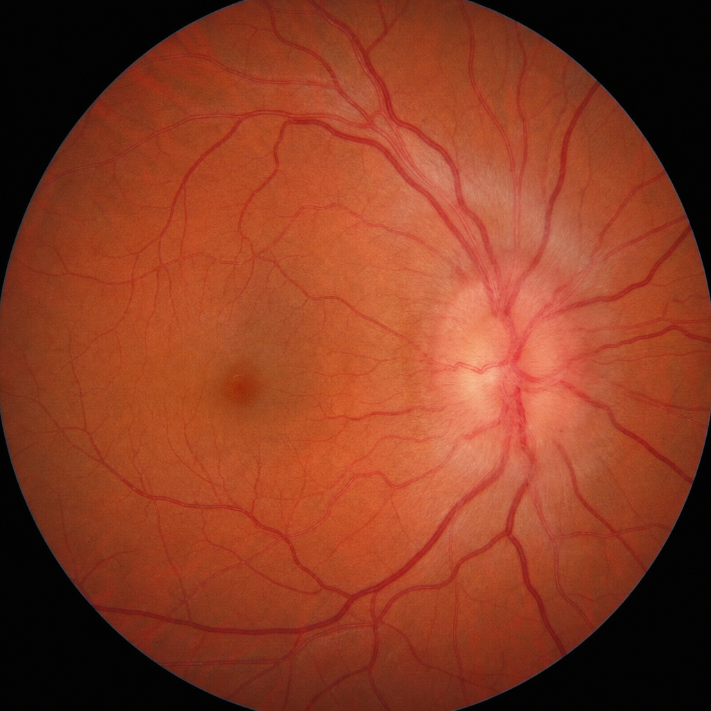
What is the most likely diagnosis based on the findings of a fundus examination showing swelling of the optic disc?
Which type of visual defects are caused by a tumor of the pituitary gland pressing upon the optic chiasm?
A 26-year-old female presents with an insidious onset of diplopia on alternate cover test, exhibiting a right hypertropia that worsens on right head tilt and left gaze. Which muscle is paralyzed?
Practice by Chapter
Anatomy of Visual Pathways
Practice Questions
Pupillary Disorders
Practice Questions
Optic Neuritis
Practice Questions
Ischemic Optic Neuropathies
Practice Questions
Other Optic Neuropathies
Practice Questions
Papilledema
Practice Questions
Cranial Nerve Palsies
Practice Questions
Nystagmus
Practice Questions
Visual Field Defects
Practice Questions
Neuro-ophthalmic Manifestations of Intracranial Lesions
Practice Questions
Functional Visual Disorders
Practice Questions
Migraine and the Eye
Practice Questions
Want unlimited practice?
Get full access to all questions, explanations, and performance tracking.
Scan to download app