
Neuro-Ophthalmology — MCQs

On this page
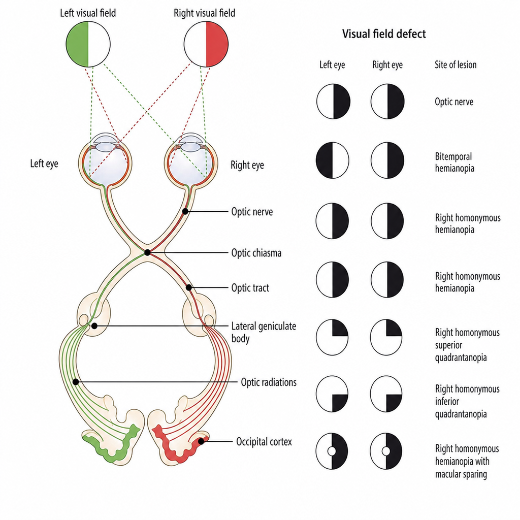
Where is a lesion that produces the visual field defect shown in the figure?
Sudden painful loss of vision is seen in which of the following conditions?
Tumours arising from all of the following structures can induce papilloedema except?
Foster Kennedy syndrome is classically described in association with which of the following tumors?
Marcus Gunn pupil is a feature of all EXCEPT:
In which of the following conditions is the retina not affected?
Which of the following is NOT true regarding Argyll Robertson pupil?
Which of the following is the drug of choice for optic neuritis?
Which cranial nerve is completely paralyzed in the case of ptosis?
What is the initial bedside investigation for papilledema?
Practice by Chapter
Anatomy of Visual Pathways
Practice Questions
Pupillary Disorders
Practice Questions
Optic Neuritis
Practice Questions
Ischemic Optic Neuropathies
Practice Questions
Other Optic Neuropathies
Practice Questions
Papilledema
Practice Questions
Cranial Nerve Palsies
Practice Questions
Nystagmus
Practice Questions
Visual Field Defects
Practice Questions
Neuro-ophthalmic Manifestations of Intracranial Lesions
Practice Questions
Functional Visual Disorders
Practice Questions
Migraine and the Eye
Practice Questions
Want unlimited practice?
Get full access to all questions, explanations, and performance tracking.
Scan to download app