A 50-year-old patient has difficulty reading close objects. Likely diagnosis?
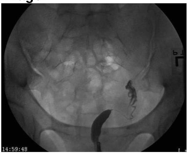
Based on the provided image, which of the following is the correct diagnosis?

In which of the following conditions is behavior therapy considered most effective?
Which of the following diseases is not included in "Vision 2020 - Right to Sight" immediate goals?
A female presents with loss of vision in the right halves of both eyes. Where is the lesion located in the optic pathway?
F00 in ICD denotes
Retinitis pigmentosa is characterized by ?
Metamorphopsia is seen in?
How can the degree of diplopia in maxillofacial trauma be accurately recorded?
Which of the following statements is MOST likely false regarding optic neuritis?
Explanation: ***Presbyopia*** - This condition is characterized by the **loss of elasticity** in the lens of the eye, which occurs naturally with age, making it difficult to focus on **near objects**. - Its typical presentation, as seen in this 50-year-old patient, is **difficulty reading close objects** or performing other tasks requiring near vision. *Hypermetropia* - Often causes **farsightedness**, meaning distant objects are seen clearly, but near objects appear blurry due to the eye attempting to constantly accommodate. - While it can make near vision difficult, it is not primarily an age-related loss of accommodation and can affect individuals of various ages. *Astigmatism* - Results from an **irregular curvature of the cornea or lens**, causing blurred or distorted vision at all distances, rather than specifically difficulty with close objects. - This condition makes it difficult for the eye to focus light uniformly on the retina, leading to multiple focal points or streaks. *Myopia* - This is commonly known as **nearsightedness**, where distant objects appear blurry while near objects are seen clearly. - It occurs when the eyeball is too long or the cornea is too steeply curved, causing light to focus in front of the retina.
Explanation: ***Unicornuate Uterus*** - The image distinctly shows **only one fallopian tube and one rudimentary uterine horn** on the right side, indicating a unicornuate uterus. - This malformation results from the **incomplete development of one Müllerian duct**, leading to a single, banana-shaped uterine cavity. *Uterus didelphys* - This condition involves **two completely separate uteri**, each with its own cervix and vagina. - The image does not show evidence of two distinct uterine bodies or cervices. *Bicornuate Uterus* - A bicornuate uterus is characterized by **two uterine horns that fuse caudally**, creating a heart-shaped appearance with a shared cervix. - The image clearly lacks the characteristic heart shape and shows only one functional horn. *Septate uterus* - A septate uterus has a **fibrous or muscular septum** dividing the uterine cavity, while the external uterine contour remains normal. - The image does not show a septum or a normal external uterine contour with an internal division; instead, it presents with a single underdeveloped horn.
Explanation: ***Obsessive-Compulsive Disorder (OCD)*** - **Exposure and Response Prevention (ERP)**, a type of behavior therapy, is the gold standard and most effective treatment for OCD. - ERP directly targets the **obsessions** and **compulsions** by gradually exposing individuals to feared situations without allowing them to perform their rituals. - OCD shows the **highest response rates** to pure behavior therapy compared to other psychiatric conditions. *Psychosis* - While supportive therapy and cognitive behavioral therapy for psychosis (CBTp) can be helpful, **behavior therapy alone is not considered the primary or most effective treatment** for core psychotic symptoms. - Management of psychosis primarily relies on **antipsychotic medications** to address symptoms like hallucinations and delusions. *Panic Attack* - Behavior therapy and CBT are effective for **Panic Disorder**, but the effectiveness is somewhat lower than for OCD. - Treatment for panic disorder often requires a **combination of behavioral and cognitive techniques** rather than pure behavior therapy alone. - Management typically includes breathing exercises, exposure to physical sensations, and cognitive restructuring. *Generalized Anxiety Disorder* - **Cognitive Behavioral Therapy (CBT)**, which includes behavioral components, is highly effective for GAD, but the **cognitive elements are essential** for addressing worry and rumination. - Pure behavior therapy (e.g., systematic desensitization) is less effective for GAD compared to OCD, as GAD involves pervasive cognitive distortions that require cognitive restructuring.
Explanation: ***Epidemic conjunctivitis*** - While a common eye infection, **epidemic conjunctivitis** is typically **self-limiting** and rarely leads to permanent blindness, thus it was not a primary target for "Vision 2020" blindness prevention programs. - The "Vision 2020" initiative focused on conditions that were the leading causes of **avoidable blindness** globally, aiming to eliminate them as public health problems. *Trachoma* - **Trachoma** is a chronic infectious eye disease that can lead to irreversible blindness and was a major focus of the "Vision 2020" initiative. - It was targeted for elimination through strategies like improved hygiene, mass drug administration, and surgical interventions. *Onchocerciasis* - Also known as **river blindness**, onchocerciasis is caused by a parasitic worm and was a significant cause of blindness in endemic regions, making it a key component of the "Vision 2020" program. - The program aimed to control and eliminate the disease through mass drug administration of ivermectin. *Cataract* - **Cataract** is the leading cause of blindness worldwide and was a primary target for the "Vision 2020" initiative due to its high prevalence and the effectiveness of surgical treatment. - The initiative aimed to increase access to affordable cataract surgery to restore sight.
Explanation: Left optic tract - A lesion in the **left optic tract** causes **right homonymous hemianopsia**, meaning loss of vision in the right halves of both eyes [1]. - This is because the left optic tract carries visual information from the **nasal retina of the right eye** and the **temporal retina of the left eye**, both of which process the right visual field [1]. *Optic radiation* - A lesion in the optic radiation would also cause a **homonymous hemianopsia** but depending on the specific location within the radiation, it could result in a **quadrantanopia** (loss of a quarter visual field) rather than a complete hemianopsia. - The optic radiation projects from the **lateral geniculate nucleus** to the **visual cortex**, and damage here affects the post-chiasmatic visual pathway [1]. *Optic chiasma* - A lesion at the **optic chiasma** typically results in **bitemporal hemianopsia**, which is the loss of vision in the **temporal halves of both eyes** [1]. - This occurs because the **crossing nasal fibers** from both eyes, which carry information from the temporal visual fields, are affected [1]. *Right optic tract* - A lesion in the **right optic tract** would result in **left homonymous hemianopsia**, meaning loss of vision in the **left halves of both eyes** [1]. - This is due to the right optic tract carrying fibers from the **nasal retina of the left eye** and the **temporal retina of the right eye**, both of which process the left visual field [1].
Explanation: ***Organic disorders - CORRECT*** - **F00-F09** in the **International Classification of Diseases (ICD-10)** Chapter V (Mental and behavioural disorders) specifically denotes **organic, including symptomatic, mental disorders** - These disorders are characterized by brain disease, brain injury, or other insult leading to **cerebral dysfunction** - **F00** specifically refers to **Dementia in Alzheimer's disease** *Mood disorders - Incorrect* - Mood disorders are classified under codes **F30-F39** in ICD-10 - This category includes conditions like bipolar affective disorder, depressive episodes, and recurrent depressive disorders *Substance use - Incorrect* - Mental and behavioral disorders due to psychoactive substance use are classified under codes **F10-F19** in ICD-10 - This section covers disorders resulting from the use of alcohol, opioids, cannabis, sedatives, hypnotics, and other substances *Psychosis - Incorrect* - Specific psychotic disorders like schizophrenia are classified under codes **F20-F29** in ICD-10 - Psychosis can be a symptom of various mental disorders, including some organic conditions
Explanation: ***Loss of peripheral vision*** - Retinitis pigmentosa is a group of genetic disorders that cause **progressive degeneration of photoreceptor cells**, primarily rods, which are concentrated in the periphery of the retina. - This degeneration leads to a gradual **loss of peripheral vision**, often beginning with **night blindness** and progressing to tunnel vision. *Loss of central vision* - Loss of central vision is characteristic of conditions affecting the **macula**, such as **age-related macular degeneration** or certain forms of **macular dystrophy**. - While later stages of retinitis pigmentosa can affect central vision, the initial and primary characteristic is loss of peripheral vision. *Tubular vision* - **Tubular vision** is an advanced stage of peripheral vision loss, where the visual field shrinks to a narrow central area, resembling looking through a tube. - While it can be a consequence of retinitis pigmentosa, it is a description of the **extent of visual field constriction** rather than the primary characteristic of the disease itself. *Visual field constriction* - **Visual field constriction** is a general term for any reduction in the extent of the visual field. - While retinitis pigmentosa certainly causes visual field constriction, the term "loss of peripheral vision" is more specific to the initial and dominant pattern of vision loss in this condition.
Explanation: ***Posterior uveitis*** - **Metamorphopsia**, or the perception of distorted images, can occur in **posterior uveitis** due to inflammation affecting the **retina** or **choroid**, leading to retinal edema or detachment. - Inflammatory processes in the posterior segment can cause disruption of photoreceptor alignment and function, altering the perception of straight lines and object shapes. *Anterior uveitis* - Primarily affects the **iris** and **ciliary body**, causing symptoms like **pain**, **photophobia**, **redness**, and reduced vision due to cellular exudates in the anterior chamber. - While vision can be affected, **metamorphopsia** is not a typical hallmark of anterior uveitis, as the retinal architecture is generally preserved. *Cataract* - Involves **clouding of the eye's natural lens**, leading to blurred vision, glare, and dullness of colors. - It does not cause **metamorphopsia** because the retinal structure and its perception of shapes remain intact; the distortion is primarily visual clarity, not shape. *Glaucoma* - Characterized by **progressive optic nerve damage** and visual field loss, often associated with elevated intraocular pressure. - While it causes vision impairment, typically starting with **peripheral vision loss**, it does not cause **metamorphopsia**, as the retina itself is usually not distorted.
Explanation: ***Hess chart*** - The **Hess chart** is a valuable tool for objectively assessing and quantifying the extent of **diplopia** by mapping the fields of gaze and identifying specific muscle palsies. - It helps in documenting the size and direction of the *deviation of the eye*, crucial for monitoring improvement or deterioration over time in **maxillofacial trauma**. *Glasgow scale* - The **Glasgow Coma Scale (GCS)** is used to assess the level of **consciousness** in patients with head injuries, not specifically for diplopia. - It evaluates eye opening, verbal response, and motor response, providing a general measure of *neurological impairment*. *Force duction test* - The **forced duction test** is a diagnostic procedure performed by the clinician to differentiate between *restrictive extraocular muscle entrapment* and *paretic muscles*. - It assesses the mechanical restriction of globe movement but does not quantify the patient's perception of **diplopia**. *None* - This option is incorrect as the **Hess chart** is a recognized and effective method for accurately recording the degree of **diplopia**.
Explanation: ***Abnormal electroretinogram*** - Optic neuritis primarily affects the **optic nerve**, which is responsible for transmitting visual information from the retina to the brain. - The **electroretinogram (ERG)** measures the electrical activity of the **retina** in response to light, which is usually normal in optic neuritis as the retina itself is not the primary site of pathology. *Decreased pupillary reflex* - Optic neuritis often causes a **relative afferent pupillary defect (RAPD)**, where the affected eye's pupil dilates instead of constricting when light is swung from the unaffected to the affected eye. - This indicates a decrease in the afferent nerve signal transmission due to damage to the optic nerve. *Decreased visual acuity* - A hallmark symptom of optic neuritis is **acute vision loss**, which can range from mild blurring to severe vision impairment. - This vision loss is typically unilateral and can progress over several days. *Abnormal visual evoked potentials (VEP)* - **VEPs** measure the electrical activity of the brain in response to visual stimuli, assessing the integrity of the optic nerve and visual pathways. - In optic neuritis, the demyelination and damage to the optic nerve cause a **slowing of nerve conduction**, leading to increased latency and reduced amplitude in VEPs.
Get full access to all questions, explanations, and performance tracking.
Start For Free