
Glaucoma — MCQs

On this page
Coloured halos are seen in all, except?
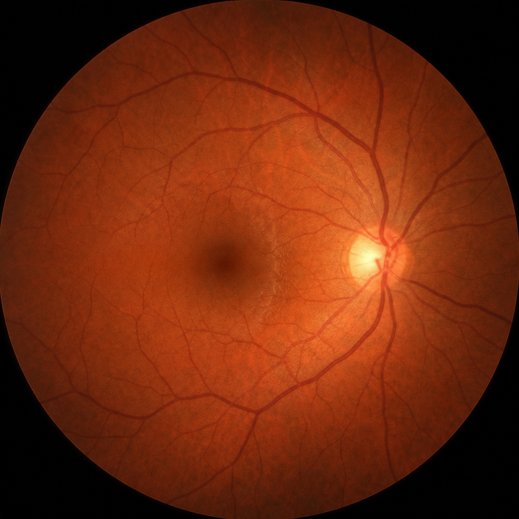
What is the diagnosis based on the following fundus examination findings?
A 60-year-old woman presents with eye pain. Her intraocular pressure is 22 mm Hg in the right eye and 25 mm Hg in the left eye. What is the most likely cause of her eye pain?
Which of the following substances is found in higher concentration in aqueous humor compared to plasma?
What is the drug of choice for primary open-angle glaucoma?
Miotics are used in the treatment of which condition?
Which drug is contraindicated in a patient with narrow-angle glaucoma?
Which of the following is true about primary angle closure glaucoma?
Which of the following is NOT a symptom of angle closure glaucoma?
A patient complains of seeing colored halos around lights in the evening and blurring of vision for the past few days, with normal intraocular pressure (IOP). What is the most likely diagnosis?
Practice by Chapter
Aqueous Humor Dynamics
Practice Questions
Primary Open-Angle Glaucoma
Practice Questions
Primary Angle-Closure Glaucoma
Practice Questions
Secondary Open-Angle Glaucomas
Practice Questions
Secondary Angle-Closure Glaucomas
Practice Questions
Developmental and Congenital Glaucomas
Practice Questions
Medical Management of Glaucoma
Practice Questions
Laser Therapy in Glaucoma
Practice Questions
Glaucoma Filtration Surgery
Practice Questions
Glaucoma Drainage Devices
Practice Questions
Angle Assessment Techniques
Practice Questions
Visual Field Testing in Glaucoma
Practice Questions
Want unlimited practice?
Get full access to all questions, explanations, and performance tracking.
Scan to download app