
Glaucoma — MCQs

On this page
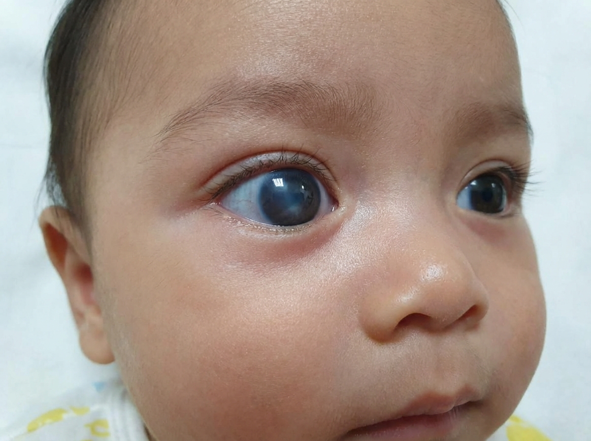
Comment on the diagnosis?
A patient presents with a miotic pupil of size 2 mm in diameter. The intraocular pressure (IOP) is 25 mm Hg. The cornea is hazy. The anterior chamber is shallow in the fellow eye. What is the most probable diagnosis?
Which of the following is NOT used in the treatment of glaucoma?
Malignant glaucoma is seen in which of the following conditions?
A patient presents with diabetic macular edema and glaucoma. Which of the following drugs should be used last for this patient?
In Fincham's test, what is observed?
Which field of vision is the last to be affected in chronic simple glaucoma?
Which of the following is NOT a mechanism in hemolytic glaucoma?
Which of the following statements regarding the optic nerve head is NOT true?
Which of the following is NOT a risk factor for primary open-angle glaucoma?
Practice by Chapter
Aqueous Humor Dynamics
Practice Questions
Primary Open-Angle Glaucoma
Practice Questions
Primary Angle-Closure Glaucoma
Practice Questions
Secondary Open-Angle Glaucomas
Practice Questions
Secondary Angle-Closure Glaucomas
Practice Questions
Developmental and Congenital Glaucomas
Practice Questions
Medical Management of Glaucoma
Practice Questions
Laser Therapy in Glaucoma
Practice Questions
Glaucoma Filtration Surgery
Practice Questions
Glaucoma Drainage Devices
Practice Questions
Angle Assessment Techniques
Practice Questions
Visual Field Testing in Glaucoma
Practice Questions
Want unlimited practice?
Get full access to all questions, explanations, and performance tracking.
Scan to download app