
Glaucoma — MCQs

On this page
Festooned (irregular) pupil is most commonly seen in which type of glaucoma?
Which of the following structures is NOT part of the trabecular outflow pathway?
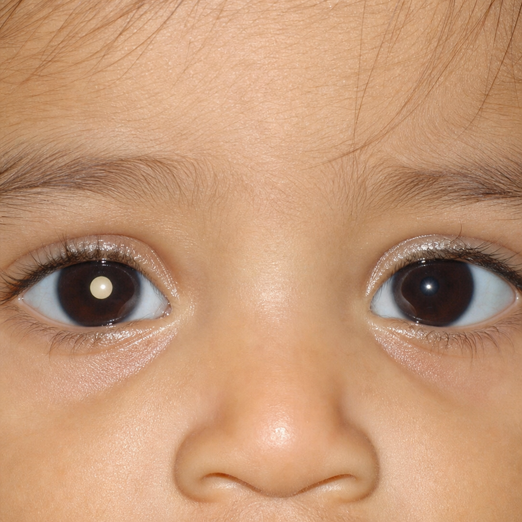
A mother brings her 18-month-old son to the paediatrician after noticing a white glow in his right eye in flash photographs over the past two months. There is no family history of eye disease. The child appears otherwise well and is reaching developmental milestones. The finding in the right eye is shown in Image 1. Which of the following is the most appropriate immediate next step in management?
A 75-year-old patient presents with deterioration of vision. On examination, the pupillary reflex is observed to be sluggish, and the intraocular pressure is normal. Optic disc evaluation shows a large and deep cup and paracentral scotomas. What is the most likely diagnosis?
What is the area of the cornea indented by a Goldmann applanation tonometer when measuring intraocular pressure?
Intractable secondary glaucoma is seen in which of the following conditions?
Pilocarpine is the drug of choice in which type of glaucoma?
A 56-year-old female presents with acute narrow-angle glaucoma, characterized by severe eye pain that radiates. In what distribution does this pain typically spread?
What is the normal aqueous production rate?
Which of the following is NOT an early sign of glaucoma?
Practice by Chapter
Aqueous Humor Dynamics
Practice Questions
Primary Open-Angle Glaucoma
Practice Questions
Primary Angle-Closure Glaucoma
Practice Questions
Secondary Open-Angle Glaucomas
Practice Questions
Secondary Angle-Closure Glaucomas
Practice Questions
Developmental and Congenital Glaucomas
Practice Questions
Medical Management of Glaucoma
Practice Questions
Laser Therapy in Glaucoma
Practice Questions
Glaucoma Filtration Surgery
Practice Questions
Glaucoma Drainage Devices
Practice Questions
Angle Assessment Techniques
Practice Questions
Visual Field Testing in Glaucoma
Practice Questions
Want unlimited practice?
Get full access to all questions, explanations, and performance tracking.
Scan to download app