Most common type of scleritis is
Most common cause of posterior staphyloma?
Recurrent anterior uveitis with increased intraocular tension is seen in which of the following conditions?
A man presents 6 hrs after head injury complaining of mild proptosis and scleral hyperemia:
Which of the following conditions is associated with granulomatous uveitis?
Most reliable sign of posterior scleritis is:
Most common type of scleritis among the following is
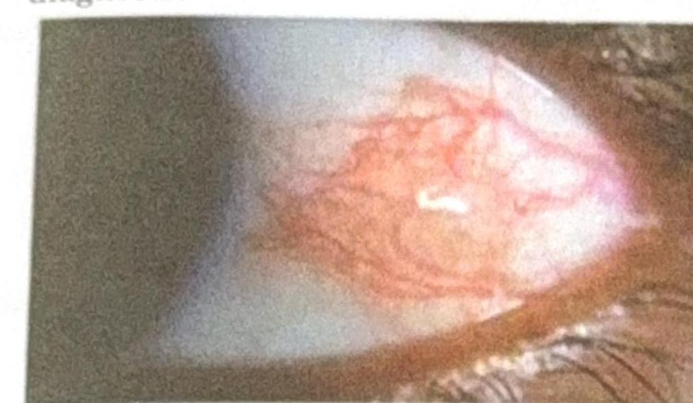
A patient presents with a nodular swelling near the limbus, which does not blanch with topical vasoconstrictors and recurs after treatment. Based on the image and clinical presentation, what is the most probable diagnosis?

Ciliary staphyloma occurs due to all of the following except:
A patient presents with proptosis, restriction of eye movements, and is found to be euthyroid. What is the most likely diagnosis?
Explanation: ***Anterior*** - **Anterior scleritis** accounts for approximately **98%** of all scleritis cases, making it the most common type. - It involves inflammation of the sclera anterior to the **equator of the globe**, visible on external examination. - Anterior scleritis is further subdivided into **diffuse, nodular,** and **necrotizing** forms based on clinical presentation and severity. *Diffuse anterior* - **Diffuse anterior scleritis** is the most common subtype of anterior scleritis, characterized by widespread inflammation. - While common among anterior types, it represents a subset rather than the overall most common anatomical category. *Nodular anterior* - **Nodular anterior scleritis** presents with discrete nodules of inflamed scleral tissue. - It is less common than diffuse anterior scleritis but more common than necrotizing forms. *Posterior* - **Posterior scleritis** is rare, accounting for only about **2%** of all scleritis cases. - It involves inflammation posterior to the **equator of the globe** and can be difficult to diagnose due to its hidden location, often presenting with pain, proptosis, and vision loss.
Explanation: ***Myopia*** - **Posterior staphyloma** is a characteristic degenerative change in **high myopia**, where the sclera thins and bulges posteriorly. - The rapid and excessive axial elongation of the eyeball in myopia leads to stretching and weakening of the posterior sclera. *Hypermetropia* - **Hypermetropia** (farsightedness) is characterized by an eyeball that is too short, leading to light focusing behind the retina. - It is not associated with the pathological thinning and bulging of the posterior sclera seen in staphyloma. *conjunctivitis* - **Conjunctivitis** is an inflammation of the conjunctiva, the membrane lining the inside of the eyelids and covering the sclera. - It does not involve structural changes to the sclera or retina that would lead to posterior staphyloma. *Glaucoma* - **Glaucoma** is a group of diseases that damage the optic nerve, often due to high intraocular pressure, leading to vision loss. - While it can cause optic disc cupping, it is not directly associated with the development of posterior staphyloma.
Explanation: ***Posner-Schlossman syndrome*** - Characterized by **recurrent, unilateral, non-granulomatous anterior uveitis** associated with markedly **elevated intraocular pressure (IOP)**. - The condition is also known as **glaucomatocyclitic crisis**, highlighting the episodic inflammation and glaucoma. - Key features include **acute attacks** lasting hours to weeks with **dramatic IOP elevation** (often >40 mmHg). *Foster-Kennedy syndrome* - This syndrome is defined by ipsilateral **optic atrophy**, contralateral **papilledema**, and often **anosmia**, typically due to a frontal lobe tumor. - It does not involve anterior uveitis or primary elevated intraocular tension. - This is a neuro-ophthalmologic syndrome, not an inflammatory ocular condition. *Vogt-Koyanagi-Harada syndrome* - An autoimmune disorder affecting pigmented tissues, leading to **bilateral granulomatous panuveitis**, often with hearing loss, vitiligo, poliosis, and neurological symptoms. - While it involves uveitis, it is typically **bilateral and panuveitis**, not recurrent unilateral anterior uveitis. - IOP may be elevated but not the defining feature with dramatic episodic rises. *Fuchs heterochromic iridocyclitis* - A chronic, **unilateral, low-grade anterior uveitis** with characteristic iris heterochromia. - May have mild IOP elevation but **not recurrent episodic attacks** with marked pressure spikes. - Inflammation is typically **quiet and chronic** rather than acute and recurrent.
Explanation: ***Retro-orbital hematoma*** - The sudden onset of **proptosis** and **scleral hyperemia** within hours of a head injury is highly suggestive of bleeding behind the eye. - A **retro-orbital hematoma** causes increased orbital pressure, leading to the forward displacement of the eyeball (proptosis) and conjunctival injection (scleral hyperemia). *Caroticocavernous fistula* - This condition involves an abnormal communication between the carotid artery and the cavernous sinus, typically presenting with a **pulsatile proptosis** and a **bruit** over the eye. - While it can cause proptosis and hyperemia, its onset is usually not as acute as 6 hours post-trauma without being a direct major vessel injury in a recent trauma. *Pneumo-orbit* - A pneumo-orbit involves **air entering the orbit**, often following trauma that fractures an orbital wall communicating with a paranasal sinus. - Symptoms include **periorbital crepitus** and exophthalmos, but scleral hyperemia is not a primary or dominant feature. *Orbital cellulitis* - Orbital cellulitis is an **infection of the orbital tissues**, usually presenting with proptosis, ophthalmoplegia, pain, and fever. - This is an infectious process and would typically take longer than 6 hours to develop to such an extent after an acute trauma without an open wound or obvious contamination.
Explanation: ***Vogt-Koyanagi-Harada's disease*** - This systemic autoimmune condition characteristically presents with **bilateral, chronic granulomatous panuveitis**. - Other typical findings include **neurologic symptoms** (meningism, tinnitus) and **dermatologic manifestations** (vitiligo, poliosis, alopecia). *Fuchs' heterochromic iridocyclitis* - This condition is typically associated with **non-granulomatous anterior uveitis**. - Key features include **heterochromia** (different colored irises), diffuse stellate keratic precipitates, and often a cataract. *Behcet's disease* - Behcet's disease causes a **non-granulomatous panuveitis**, which is often recurrent and severe. - It is characterized by **recurrent oral and genital ulcers**, erythema nodosum, and pathergy. *Psoriatic arthritis* - Uveitis associated with psoriatic arthritis is usually **acute, unilateral, and non-granulomatous anterior uveitis**. - It is often seen in conjunction with **psoriatic skin lesions** and arthritis, commonly affecting the peripheral joints.
Explanation: ***T-sign on ultrasound*** - The **T-sign** on B-scan ultrasound is considered the most reliable diagnostic sign of **posterior scleritis**. - It represents accumulation of **fluid in Tenon's capsule** and is highly specific for the condition. *Disc edema* - While **disc edema** can be a clinical finding in posterior scleritis due to inflammation, it is not specific and can be caused by various other conditions, such as **optic neuritis** or papilledema. - It is not as pathognomonic as the T-sign on ultrasound for confirming the diagnosis. *Choroidal folds* - **Choroidal folds** can occur in posterior scleritis due to scleral inflammation and thickening, which can indent the choroid. - However, choroidal folds can also be seen in other conditions like **orbital tumors** or **hypotony**, making them a less specific indicator. *Exudative detachment* - An **exudative retinal detachment** can occur in severe cases of posterior scleritis due to inflammation and fluid leakage from the inflamed choroid into the subretinal space. - This is a serious complication and a sign of advanced disease, but not the earliest or most reliable diagnostic sign for the condition itself, as it can also be seen in other inflammatory or vascular conditions.
Explanation: ***Non-necrotizing*** - This category, particularly **diffuse non-necrotizing scleritis**, is the most frequently encountered type. - It is often characterized by widespread inflammation of the sclera without tissue loss. *Necrotizing* - This is a severe form of scleritis associated with significant **tissue destruction** and a high risk of vision loss. - While serious, it is thankfully much rarer than the non-necrotizing forms. *Granulomatous* - This term describes a **histopathological feature** of inflammation rather than a distinct clinical type of scleritis. - Granulomatous inflammation can be seen in various forms of scleritis but is not a primary classification of its most common presentation. *Posterior* - **Posterior scleritis** specifically involves inflammation of the sclera behind the equator of the globe. - It is less common than anterior scleritis and can present with different symptoms such as vision loss or pain with eye movement.
Explanation: ***Scleritis with rheumatoid arthritis*** - The image shows **deep, violaceous conjunctival injection** with surrounding edema and a nodular appearance near the limbus, consistent with **nodular scleritis**. Scleritis is inflammation of the sclera, often characterized by severe pain and association with systemic autoimmune diseases like **rheumatoid arthritis**, which can cause destructive lesions and recurrence. - The characteristic **deep vessel engorgement that does not blanch with phenylephrine** and the history of recurrence further support scleritis. *Episcleritis with rheumatoid arthritis* - Episcleritis presents with a **more superficial, bright red injection** involving the episclera, which generally **blanches with topical phenylephrine** and is less painful than scleritis. - While episcleritis can be associated with rheumatoid arthritis, the clinical features described (nodular swelling, deep injection) are more typical of scleritis. *Pinguecula* - A pinguecula is a **yellowish patch or bump** on the conjunctiva, typically on the nasal side of the eye, that is **not inflamed** unless irritated. - It consists of **degenerated collagen fibers** and elastic tissue, and does not present with the diffuse, deep vascular injection seen in the image. *Dry eye* - Dry eye is characterized by **ocular dryness, irritation, and sometimes a foreign body sensation**, but it typically causes **diffuse conjunctival hyperemia** rather than a localized, nodular, deep inflammation with surrounding edema as shown. - While dry eye can be associated with autoimmune diseases, its appearance is not consistent with the depicted nodular lesion.
Explanation: ***Episcleritis*** - **Episcleritis** is a benign, self-limiting inflammation of the episclera, which is a superficial layer of connective tissue, and does not lead to **scleral thinning** or ectasia. - Therefore, it does not cause **ciliary staphyloma**, which is a bulging of the sclera in the ciliary body region. *Scleritis* - **Scleritis** is a severe, chronic inflammatory disease affecting the sclera, often leading to **scleral thinning** and weakening. - This thinning can predispose to the formation of a **ciliary staphyloma**, especially if the inflammation is localized in the ciliary region. *Absolute glaucoma* - **Absolute glaucoma** is a severe form of glaucoma characterized by persistently high **intraocular pressure (IOP)**, leading to total vision loss and often significant **scleral thinning** due to chronic pressure. - The elevated IOP can cause stretching and thinning of the sclera, particularly in weakened areas like the **ciliary body**, resulting in a **ciliary staphyloma**. *Perforating injury* - A **perforating injury** to the globe can directly weaken the **scleral wall**, especially if it occurs near the **ciliary body**. - Subsequent healing with scar tissue, often under intraocular pressure, can lead to ectasia and the formation of a **ciliary staphyloma**.
Explanation: ***Orbital pseudotumor*** - **Orbital pseudotumor** (also known as idiopathic orbital inflammation) presents with **proptosis** and **restriction of eye movements**. - Typically presents with **acute or subacute onset** of **painful** ophthalmoplegia and proptosis. - It is a **diagnosis of exclusion** after ruling out other causes of orbital inflammation, including thyroid eye disease, orbital cellulitis, and orbital neoplasms. - **Euthyroid status** does not exclude this diagnosis, and it is the **most common painful orbital mass** in adults. *Orbital cellulitis* - **Orbital cellulitis** presents with **rapid onset** of **painful proptosis**, **chemosis**, **ophthalmoplegia**, and signs of **acute infection** (fever, periorbital erythema, recent sinusitis). - The absence of infectious signs and acute inflammatory markers makes this less likely. *Orbital lymphoma* - **Orbital lymphoma** typically presents with **slowly progressive, painless proptosis** in older patients. - It is a **chronic, indolent process** and less likely to cause acute, painful restriction of eye movements. - Usually presents as a palpable mass in the superotemporal orbit. *Thyroid eye disease* - **Thyroid eye disease** (Graves' ophthalmopathy) commonly causes **proptosis** and **restricted eye movements** due to extraocular muscle enlargement. - While **90% of TED patients have hyperthyroidism**, approximately **5% are euthyroid at presentation** (euthyroid Graves' ophthalmopathy). - However, TED typically has a **subacute to chronic onset**, bilateral involvement, lid retraction, and characteristic imaging findings (muscle belly enlargement sparing tendons). - The clinical presentation with acute symptoms and euthyroid state makes **orbital pseudotumor more likely** as the initial diagnosis.
Get full access to all questions, explanations, and performance tracking.
Start For Free