Recurrent anterior uveitis with increased intraocular tension is seen in which of the following conditions?
In a patient with acute anterior uveitis presenting with raised intraocular pressure, the PRIMARY treatment should be:
A 40-year-old woman with rheumatoid arthritis presents with eye pain and redness. Examination reveals scleritis. What is the most likely associated systemic complication?

Most common type of scleritis is
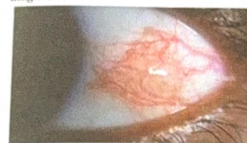
A patient presents with a nodular swelling near the limbus, which does not blanch with topical vasoconstrictors and recurs after treatment. Based on the image and clinical presentation, what is the most probable diagnosis?

Ciliary staphyloma occurs due to all of the following except:
Massaging of nasolacrimal duct is done in ?
Ciliary staphyloma is seen in which of the following conditions?
Posterior staphyloma is associated with which of the following conditions?
Sclera is thinnest at:
Explanation: ***Posner-Schlossman syndrome*** - Characterized by **recurrent, unilateral, non-granulomatous anterior uveitis** associated with markedly **elevated intraocular pressure (IOP)**. - The condition is also known as **glaucomatocyclitic crisis**, highlighting the episodic inflammation and glaucoma. - Key features include **acute attacks** lasting hours to weeks with **dramatic IOP elevation** (often >40 mmHg). *Foster-Kennedy syndrome* - This syndrome is defined by ipsilateral **optic atrophy**, contralateral **papilledema**, and often **anosmia**, typically due to a frontal lobe tumor. - It does not involve anterior uveitis or primary elevated intraocular tension. - This is a neuro-ophthalmologic syndrome, not an inflammatory ocular condition. *Vogt-Koyanagi-Harada syndrome* - An autoimmune disorder affecting pigmented tissues, leading to **bilateral granulomatous panuveitis**, often with hearing loss, vitiligo, poliosis, and neurological symptoms. - While it involves uveitis, it is typically **bilateral and panuveitis**, not recurrent unilateral anterior uveitis. - IOP may be elevated but not the defining feature with dramatic episodic rises. *Fuchs heterochromic iridocyclitis* - A chronic, **unilateral, low-grade anterior uveitis** with characteristic iris heterochromia. - May have mild IOP elevation but **not recurrent episodic attacks** with marked pressure spikes. - Inflammation is typically **quiet and chronic** rather than acute and recurrent.
Explanation: ***Topical steroids*** - **Topical corticosteroids** are the primary treatment for **anterior uveitis** to reduce inflammation, which is the underlying cause of both the uveitis and often the raised IOP. - While IOP is elevated, managing the inflammation with steroids is crucial, as the inflammation itself can lead to secondary **IOP elevation** due to trabecular meshwork dysfunction or synechiae formation. *Topical beta-blockers* - **Topical beta-blockers** are used to lower intraocular pressure, but they do not address the underlying **inflammation** in acute anterior uveitis. - Using them alone without treating the inflammation can lead to progression of the uveitis and further ocular damage. *Cycloplegics* - **Cycloplegics** (e.g., atropine, cyclopentolate) are important adjuncts in acute anterior uveitis to relieve pain from ciliary spasm and prevent posterior synechiae formation by dilating the pupil. - They do not, however, treat the **inflammation** directly or primarily address the elevated intraocular pressure. *Miotics* - **Miotics** (e.g., pilocarpine) **constrict the pupil**, which can worsen symptoms in acute anterior uveitis by increasing ciliary body spasm and potentially increasing the risk of posterior synechiae formation. - They are contraindicated in acute anterior uveitis as they exacerbate pain and inflammation, and do not treat the underlying cause.
Explanation: ***Interstitial lung disease*** - **Scleritis** in a patient with **rheumatoid arthritis** often indicates a more severe, systemic form of the disease. - **Interstitial lung disease (ILD)** is a common and serious **extra-articular manifestation** of rheumatoid arthritis, associated with higher disease activity and worse prognosis. *Cardiomyopathy* - While cardiac involvement can occur in RA (e.g., pericarditis, myocarditis), **cardiomyopathy** is less directly correlated with scleritis as a linked severe systemic complication. - **Rheumatoid arthritis** can increase the risk of cardiovascular disease due to accelerated atherosclerosis and systemic inflammation, but cardiomyopathy itself is not typically the direct systemic complication associated with scleritis. *Renal failure* - **Renal involvement** in rheumatoid arthritis is uncommon, usually related to complications from treatment (e.g., NSAID-induced nephropathy) or secondary amyloidosis, not directly with scleritis. - Scleritis itself does not directly predispose to **renal failure** as a primary systemic complication of RA. *Peripheral neuropathy* - **Peripheral neuropathy** can be a complication of rheumatoid arthritis, particularly in severe cases or vasculitis. - However, it is not as strongly or directly linked to the presence of **scleritis** as **interstitial lung disease** is, in terms of signifying systemic disease severity.
Explanation: ***Anterior*** - **Anterior scleritis** accounts for approximately **98%** of all scleritis cases, making it the most common type. - It involves inflammation of the sclera anterior to the **equator of the globe**, visible on external examination. - Anterior scleritis is further subdivided into **diffuse, nodular,** and **necrotizing** forms based on clinical presentation and severity. *Diffuse anterior* - **Diffuse anterior scleritis** is the most common subtype of anterior scleritis, characterized by widespread inflammation. - While common among anterior types, it represents a subset rather than the overall most common anatomical category. *Nodular anterior* - **Nodular anterior scleritis** presents with discrete nodules of inflamed scleral tissue. - It is less common than diffuse anterior scleritis but more common than necrotizing forms. *Posterior* - **Posterior scleritis** is rare, accounting for only about **2%** of all scleritis cases. - It involves inflammation posterior to the **equator of the globe** and can be difficult to diagnose due to its hidden location, often presenting with pain, proptosis, and vision loss.
Explanation: ***Scleritis with rheumatoid arthritis*** - The image shows **deep, violaceous conjunctival injection** with surrounding edema and a nodular appearance near the limbus, consistent with **nodular scleritis**. Scleritis is inflammation of the sclera, often characterized by severe pain and association with systemic autoimmune diseases like **rheumatoid arthritis**, which can cause destructive lesions and recurrence. - The characteristic **deep vessel engorgement that does not blanch with phenylephrine** and the history of recurrence further support scleritis. *Episcleritis with rheumatoid arthritis* - Episcleritis presents with a **more superficial, bright red injection** involving the episclera, which generally **blanches with topical phenylephrine** and is less painful than scleritis. - While episcleritis can be associated with rheumatoid arthritis, the clinical features described (nodular swelling, deep injection) are more typical of scleritis. *Pinguecula* - A pinguecula is a **yellowish patch or bump** on the conjunctiva, typically on the nasal side of the eye, that is **not inflamed** unless irritated. - It consists of **degenerated collagen fibers** and elastic tissue, and does not present with the diffuse, deep vascular injection seen in the image. *Dry eye* - Dry eye is characterized by **ocular dryness, irritation, and sometimes a foreign body sensation**, but it typically causes **diffuse conjunctival hyperemia** rather than a localized, nodular, deep inflammation with surrounding edema as shown. - While dry eye can be associated with autoimmune diseases, its appearance is not consistent with the depicted nodular lesion.
Explanation: ***Episcleritis*** - **Episcleritis** is a benign, self-limiting inflammation of the episclera, which is a superficial layer of connective tissue, and does not lead to **scleral thinning** or ectasia. - Therefore, it does not cause **ciliary staphyloma**, which is a bulging of the sclera in the ciliary body region. *Scleritis* - **Scleritis** is a severe, chronic inflammatory disease affecting the sclera, often leading to **scleral thinning** and weakening. - This thinning can predispose to the formation of a **ciliary staphyloma**, especially if the inflammation is localized in the ciliary region. *Absolute glaucoma* - **Absolute glaucoma** is a severe form of glaucoma characterized by persistently high **intraocular pressure (IOP)**, leading to total vision loss and often significant **scleral thinning** due to chronic pressure. - The elevated IOP can cause stretching and thinning of the sclera, particularly in weakened areas like the **ciliary body**, resulting in a **ciliary staphyloma**. *Perforating injury* - A **perforating injury** to the globe can directly weaken the **scleral wall**, especially if it occurs near the **ciliary body**. - Subsequent healing with scar tissue, often under intraocular pressure, can lead to ectasia and the formation of a **ciliary staphyloma**.
Explanation: ***Congenital dacryocystitis*** - **Massaging the nasolacrimal duct** (Crigler massage) is a primary treatment for congenital dacryocystitis to promote the opening of the **valve of Hasner**. - This condition is due to incomplete canalization of the nasolacrimal duct, leading to tearing and discharge in infants. *Acute dacryocystitis* - This is an **acute infection of the lacrimal sac**, and massaging can worsen the condition by spreading the infection. - Treatment typically involves **antibiotics** and, if necessary, incision and drainage of any abscess. *Conjunctivitis* - **Conjunctivitis** is inflammation of the conjunctiva and is not related to obstruction of the nasolacrimal duct. - Massaging the nasolacrimal duct has no therapeutic role in treating conjunctivitis. *None of the options* - This option is incorrect because **congenital dacryocystitis** is a condition where nasolacrimal duct massage is a standard and effective treatment.
Explanation: **Explanation:** **Ciliary staphyloma** is a localized thinning and bulging of the sclera lined by the underlying ciliary body. It occurs due to a combination of weakened scleral integrity and chronically elevated intraocular pressure (IOP). 1. **Why Absolute Glaucoma is correct:** In absolute glaucoma, the IOP is severely and chronically elevated. This persistent pressure causes the sclera to stretch and thin, particularly in the **ciliary zone** (the area 2–8 mm behind the limbus). As the sclera thins, the dark pigment of the underlying ciliary body shines through, giving it a characteristic bluish-black appearance. 2. **Why the other options are incorrect:** * **Pathological Myopia:** This is typically associated with **Posterior staphyloma**, where the thinning occurs at the posterior pole (macular area) due to excessive axial elongation. * **Retinoblastoma:** While advanced tumors can cause globe enlargement (buphthalmos) or extraocular extension, they do not typically present as a focal ciliary staphyloma. * **Episcleritis:** This is a self-limiting, superficial inflammation of the episcleral tissues that does not lead to scleral thinning or staphyloma formation. **Clinical Pearls for NEET-PG:** * **Types of Staphyloma:** * **Anterior (Intercalary):** At the limbus; seen in secondary glaucoma following corneal perforation. * **Ciliary:** 2–8 mm from limbus; seen in Absolute Glaucoma and Scleritis. * **Equatorial:** At the exit of vortex veins; seen in Pathological Myopia. * **Posterior:** At the posterior pole; hallmark of Pathological Myopia. * **Key Sign:** The "bluish" color of a staphyloma is not due to the sclera itself, but the **uveal tissue** visible through the thinned sclera.
Explanation: **Explanation:** **Posterior staphyloma** is defined as a localized bulging of the weak, thinned sclera lined by uveal tissue, occurring posterior to the equator of the eyeball. **1. Why Pathological Myopia is correct:** In **Pathological (High) Myopia**, there is progressive axial elongation of the eyeball. This leads to mechanical stretching and thinning of the posterior pole of the sclera. As the sclera weakens, it bulges outward, creating a staphyloma. This is a hallmark feature of pathological myopia and is often associated with "lacquer cracks," chorioretinal atrophy, and Foster-Fuchs spots. **2. Why the other options are incorrect:** * **Uveoscleritis:** While inflammation can weaken the sclera, it typically leads to generalized thinning or anterior/ciliary staphylomas rather than the classic posterior staphyloma. * **Pseudocornea:** This refers to a layer of organized exudates and fibrous tissue covering a perforated cornea. It is a feature of anterior segment pathology, not posterior scleral ectasia. * **Angle-closure glaucoma:** Chronic high intraocular pressure in children (Buphthalmos) can lead to generalized enlargement of the globe, but in adults, glaucoma does not typically cause posterior staphyloma. **Clinical Pearls for NEET-PG:** * **Types of Staphyloma:** * **Anterior:** At the limbus (due to perforated corneal ulcer). * **Ciliary:** Over the ciliary body (2–3 mm behind the limbus). * **Equatorial:** At the equator (where vortex veins exit). * **Posterior:** At the posterior pole (Pathognomonic for Pathological Myopia). * **Diagnosis:** Posterior staphyloma is best visualized using **B-scan Ultrasonography** or MRI. * **Key association:** It is the most common cause of a "long" axial length on biometry.
Explanation: The sclera is the dense, fibrous outer protective coat of the eye. Its thickness varies significantly across different anatomical zones, which is a high-yield concept for surgical and clinical ophthalmology. ### **Explanation of the Correct Answer** **D. Points of muscular attachments:** The sclera is thinnest at the insertion points of the **extraocular muscles (EOMs)**, specifically just posterior to their tendons. At these sites, the scleral thickness is approximately **0.3 mm**. This anatomical vulnerability is clinically significant during strabismus surgery, as there is a higher risk of scleral perforation during muscle recession or resection. ### **Analysis of Incorrect Options** * **A. Posterior pole:** This is the **thickest** part of the sclera, measuring approximately **1.0 mm to 1.3 mm**. The thickness here provides structural support near the optic nerve head. * **B. Equator:** At the equator, the sclera has an intermediate thickness of about **0.4 mm to 0.6 mm**. * **C. Corneoscleral junction (Limbus):** The sclera is relatively thick here, measuring approximately **0.8 mm**. ### **NEET-PG High-Yield Pearls** * **Thickness Gradient:** Posterior pole (1.0 mm) → Limbus (0.8 mm) → Equator (0.5 mm) → Muscle Insertions (0.3 mm). * **Composition:** The sclera is primarily composed of Type I collagen. It is relatively avascular, receiving its nutrition from the episclera and underlying choroid. * **Lamina Cribrosa:** This is a sieve-like portion of the sclera at the optic nerve head; it is the weakest point of the outer coat against intraocular pressure (relevant in glaucoma). * **Scleral Blue Discoloration:** Seen in conditions where the sclera thins (e.g., high myopia, scleromalacia perforans) or in systemic diseases like Osteogenesis Imperfecta (due to Type I collagen defect).
Get full access to all questions, explanations, and performance tracking.
Start For Free