Most common cause of posterior staphyloma?
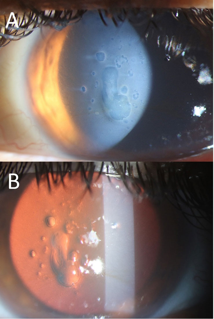
A patient, three years post-surgery, presents with the condition shown in the image and complains of decreased vision. What is the most likely diagnosis?

Which of the following is NOT a connective tissue disorder?
A 10-year-old boy presents with frequent fractures and blue sclerae. This patient most likely carries a mutation in a gene that encodes which of the following proteins?
Osteogenesis imperfecta is due to a defect in what?
Blue sclera is associated with:
Blue sclera is seen in-
Blue sclera is seen in all of the following conditions except:
An intrauterine scan at the 13th week of pregnancy showed a fetus with multiple long bone fractures. What is commonly associated with this finding?
A 22-year-old Air-force test pilot presents after flying a sortie. He reports no pain or vision changes. Eye examination reveals a localized red patch on the sclera. What is the most likely diagnosis?
Explanation: ***Myopia*** - **Posterior staphyloma** is a characteristic degenerative change in **high myopia**, where the sclera thins and bulges posteriorly. - The rapid and excessive axial elongation of the eyeball in myopia leads to stretching and weakening of the posterior sclera. *Hypermetropia* - **Hypermetropia** (farsightedness) is characterized by an eyeball that is too short, leading to light focusing behind the retina. - It is not associated with the pathological thinning and bulging of the posterior sclera seen in staphyloma. *conjunctivitis* - **Conjunctivitis** is an inflammation of the conjunctiva, the membrane lining the inside of the eyelids and covering the sclera. - It does not involve structural changes to the sclera or retina that would lead to posterior staphyloma. *Glaucoma* - **Glaucoma** is a group of diseases that damage the optic nerve, often due to high intraocular pressure, leading to vision loss. - While it can cause optic disc cupping, it is not directly associated with the development of posterior staphyloma.
Explanation: ***Posterior Capsular Opacification*** - The image (especially part B, the **red reflex view**) shows a cloudy membrane with **Elschnig pearls** and fibrous tissue behind the intraocular lens, which is characteristic of **PCO**. - PCO commonly occurs months to years after cataract surgery and causes **decreased vision** due to scattering of light. *Corneal Dystrophy* - Corneal dystrophies primarily affect the **cornea**, leading to various forms of corneal opacification, and would typically appear as cloudiness or irregularities within the corneal layers. - While it can cause decreased vision, the opacification seen in the image is clearly behind the iris plane and involves the posterior capsule, not the cornea. *Pseudophakic Bullous Keratopathy* - This condition involves **corneal edema** and **bullae formation** due to endothelial cell dysfunction, leading to a hazy cornea. - The image does not show corneal edema or bullae; instead, the primary pathology is located at the **posterior capsule** of the intraocular lens. *Posterior Subcapsular Cataract (PSC)* - A PSC is a type of cataract that forms **before cataract surgery** and affects the natural crystalline lens. - The patient is three years post-surgery, indicating that the natural lens has been replaced, making a PSC (of the natural lens) an impossible diagnosis in this context.
Explanation: ***Sjogren's syndrome*** - Sjogren's syndrome is an **autoimmune disease** characterized by **dry eyes** and **dry mouth**, resulting from immune-mediated destruction of exocrine glands [1]. - While it can involve multiple organ systems and is classified as a **systemic autoimmune disorder**, it is primarily an **exocrinopathy**, and not exclusively a disorder of connective tissue structure or function. *Osteoarthritis* - Osteoarthritis is a **degenerative joint disease** primarily affecting the **articular cartilage** and underlying bone. - While it involves destruction of cartilage, which is a connective tissue, it is considered a **degenerative rather than a systemic inflammatory or autoimmune connective tissue disorder**. *Fibromyalgia* - Fibromyalgia is a chronic pain condition characterized by **widespread musculoskeletal pain**, fatigue, and sleep disturbances, without evidence of inflammation or tissue damage [3]. - It is considered a **central sensitization syndrome** rather than a disorder of the connective tissue itself [3]. *SLE* - Systemic lupus erythematosus (SLE) is a **prototypical systemic autoimmune connective tissue disorder** affecting multiple organ systems [2]. - It is characterized by **autoantibody production** and immune complex deposition, leading to inflammation and damage in joints, skin, kidneys, and other tissues [2].
Explanation: ***Collagen*** - Collagen mutations commonly result in various **connective tissue disorders** and defects in structural integrity [1]. - It plays a critical role in **skin, bones, and cartilage**, making it a significant candidate for mutations affecting these areas [1]. *Keratin* - Keratin is a protein primarily found in **skin, hair, and nails**, and mutations typically cause **epidermal dysplasia or disorders** like keratoderma. - While important for structural integrity, keratin is less associated with broader connective tissue disorders. *Fibrillin* - Fibrillin is essential for maintaining the **elasticity and structure of connective tissues**, predominantly in **Marfan syndrome** [2]. - Mutations lead to issues with connective tissue but are not as broadly relevant as collagen in this context [2]. *Myosin* - Myosin is primarily involved in **muscle contraction** and is not typically associated with **connective tissue** disorders. - Mutations in myosin often relate to **muscle diseases**, which are not relevant to the context of connective tissue mutations. **References:** [1] Kumar V, Abbas AK, et al.. Robbins and Cotran Pathologic Basis of Disease. 9th ed. Genetic Disorders, pp. 154-155. [2] Kumar V, Abbas AK, et al.. Robbins and Cotran Pathologic Basis of Disease. 9th ed. Genetic Disorders, pp. 153-154.
Explanation: ***Collagen 1*** - Osteogenesis imperfecta is primarily caused by a defect in **type I collagen** [2], which is crucial for bone strength and structure. - This defect leads to **brittle bones**, resulting in frequent fractures and skeletal deformities . *Collagen 2* - Type II collagen is mainly found in **cartilage** and is critical for **hyaline cartilage formation**, not directly involved in bone integrity. - Defects in type II collagen are associated with conditions like **chondrodysplasia**, rather than osteogenesis imperfecta. *Collagen 4* - Type IV collagen is primarily found in **basement membranes** and plays a role in filtration and structural integrity of tissues. - While important for kidney and eye function, it is not related to the bone fragility seen in osteogenesis imperfecta. *Collagen 3* - Type III collagen is involved in the structure of **reticular fibers** and is crucial for skin and blood vessel integrity. - It is not the primary collagen affected in osteogenesis imperfecta, which is associated specifically with type I collagen defects. **References:** [1] Kumar V, Abbas AK, et al.. Robbins and Cotran Pathologic Basis of Disease. 9th ed. Bones, Joints, and Soft Tissue Tumors, p. 1182. [2] Kumar V, Abbas AK, et al.. Robbins and Cotran Pathologic Basis of Disease. 9th ed. Bones, Joints, and Soft Tissue Tumors, p. 1188.
Explanation: ***Osteogenesis imperfecta*** - **Osteogenesis imperfecta** (brittle bone disease) is the **MOST CLASSIC** condition associated with blue sclera. - Caused by a defect in **Type I collagen synthesis**, which is a major structural component of the sclera. - The deficiency in functional collagen leads to an **abnormally thin and translucent sclera**, allowing the underlying **choroidal pigment** to show through, giving it a characteristic blue appearance. - Blue sclera is seen in **80-90% of patients** with Type I osteogenesis imperfecta. *Buphthalmos* - **Buphthalmos** (congenital glaucoma) can occasionally show bluish sclera due to scleral thinning from increased intraocular pressure. - However, the primary findings are **corneal enlargement, corneal edema**, and increased eye size, not blue sclera as a defining feature. *Marfan's syndrome* - **Marfan's syndrome** is a connective tissue disorder caused by **fibrillin-1** defects. - Classic ocular features include **ectopia lentis** (lens dislocation), high myopia, and retinal detachment risk. - Blue sclera is **NOT a recognized clinical feature** of Marfan's syndrome. *Ehlers-Danlos syndrome* - This connective tissue disorder can present with blue sclera due to collagen abnormalities. - However, it is **less common** than osteogenesis imperfecta as a cause of blue sclera.
Explanation: Blue sclera is seen in- ***Osteogenesis imperfecta*** - **Blue sclera** is a classic sign due to the underlying **collagen disorder** where the sclera is thinner, allowing the underlying choroidal vessels to show through. - This genetic disorder is characterized by **brittle bones** and **connective tissue fragility**. *Ehlers-Danlos syndrome* - While it is a **collagen disorder** and can cause **connective tissue laxity**, **blue sclera** is not a primary or common feature. [1] - It often presents with **hyperextensible joints**, **fragile skin**, and easy bruising. [1] *Marfan syndrome* - This is a genetic disorder affecting connective tissue, primarily due to a defect in **fibrillin-1**, but it is not typically associated with **blue sclera**. - Key features include **tall stature**, **long slender limbs (arachnodactyly)**, and **cardiovascular abnormalities** like aortic dissection. *Rheumatoid arthritis* - This is an **autoimmune inflammatory disease** primarily affecting synovial joints and is not associated with **blue sclera**. - Ocular manifestations in rheumatoid arthritis typically involve **scleritis** or **episcleritis**, which are inflammatory conditions of the sclera, not a blue discoloration.
Explanation: ***Keratoconus*** - Keratoconus is a progressive eye disease in which the normally round cornea thins and begins to bulge into a cone-like shape, leading to **vision distortion**. - Blue sclera is **not a feature** of keratoconus. This is a **corneal condition** that does not affect the sclera. - Blue sclera, seen in the other conditions listed, occurs due to thinning of the sclera, making the underlying choroidal pigment visible. *Marfan's syndrome* - Patients with Marfan's syndrome can have blue sclera due to the **thinning of collagen** in the scleral tissue, allowing the underlying choroid to show through. - This connective tissue disorder affects multiple body systems, including the skeletal, cardiovascular, and ocular systems, with features like **arachnodactyly** and **aortic root dilation**. *Osteogenesis imperfecta* - Often referred to as **brittle bone disease**, osteogenesis imperfecta is characterized by defective **type I collagen synthesis**, which also affects the sclera. - The sclera becomes thin and translucent, revealing the underlying choroidal pigment, thus appearing **blue**. *Rheumatoid arthritis* - In rheumatoid arthritis, particularly with severe or long-standing disease, the sclera can become thinned due to **scleritis** or **scleromalacia perforans**. - This thinning can lead to a **blue discoloration** of the sclera, making the underlying choroid visible.
Explanation: ***Osteogenesis imperfecta*** - **Multiple long bone fractures** detected early in pregnancy are a classic presentation of **osteogenesis imperfecta (OI)**, a genetic disorder characterized by **bone fragility**. - OI is primarily caused by mutations in genes encoding **type I collagen**, leading to defective bone formation. *Achondroplasia* - This condition is a form of **dwarfism** characterized by disproportionately short limbs and a normal-sized trunk, resulting from a mutation in the **FGFR3 gene**. - While it affects bone growth, it typically does not cause **multiple fractures** prenatally. *Marfan syndrome* - This is a connective tissue disorder affecting the skeletal, ocular, and cardiovascular systems, characterized by **tall stature**, **long limbs and fingers**, and **aortic root dilation**. - It results from a mutation in the **fibrillin-1 gene** and is not primarily associated with prenatal long bone fractures. *Cretinism* - This is a historical term for **congenital hypothyroidism**, which results from severely deficient thyroid hormone production in a newborn. - It leads to developmental delays, growth retardation, and intellectual disability, but not to **multiple bone fractures**.
Explanation: ***Subconjunctival hemorrhage*** - A **localized red patch on the sclera** with no pain or vision changes, especially after activities that can increase venous pressure (like flying a sortie or straining), is characteristic of a **subconjunctival hemorrhage**. - It results from the rupture of small blood vessels beneath the conjunctiva, causing blood to pool. *Hyphema* - This involves blood in the **anterior chamber of the eye**, usually visible as a fluid level and often causing pain or blurred vision. - It typically results from **trauma** and is not described as a localized red patch on the sclera. *Keratitis* - **Keratitis** is inflammation of the cornea, causing pain, redness, photophobia, and often blurred vision. - The patient has no pain or vision changes, and the presentation is a localized scleral patch, not diffuse corneal involvement. *Allergic conjunctivitis* - Presents with **redness**, itching, tearing, and often bilateral involvement, sometimes with discharge. - The description of a solitary, localized red patch without other allergic symptoms makes this diagnosis unlikely.
Get full access to all questions, explanations, and performance tracking.
Start For Free