Type I collagen is present in all EXCEPT:
An example of a scleroprotein is
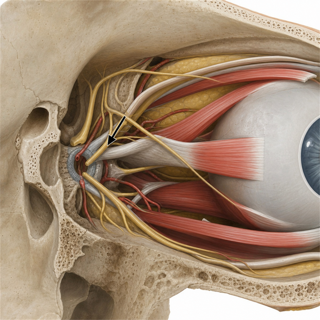
Normal intraocular pressure is typically in the range of:
Blue sclera is associated with:
A patient presents with a nodular swelling near the limbus, which does not blanch with topical vasoconstrictors and recurs after treatment. Based on the image and clinical presentation, what is the most probable diagnosis?

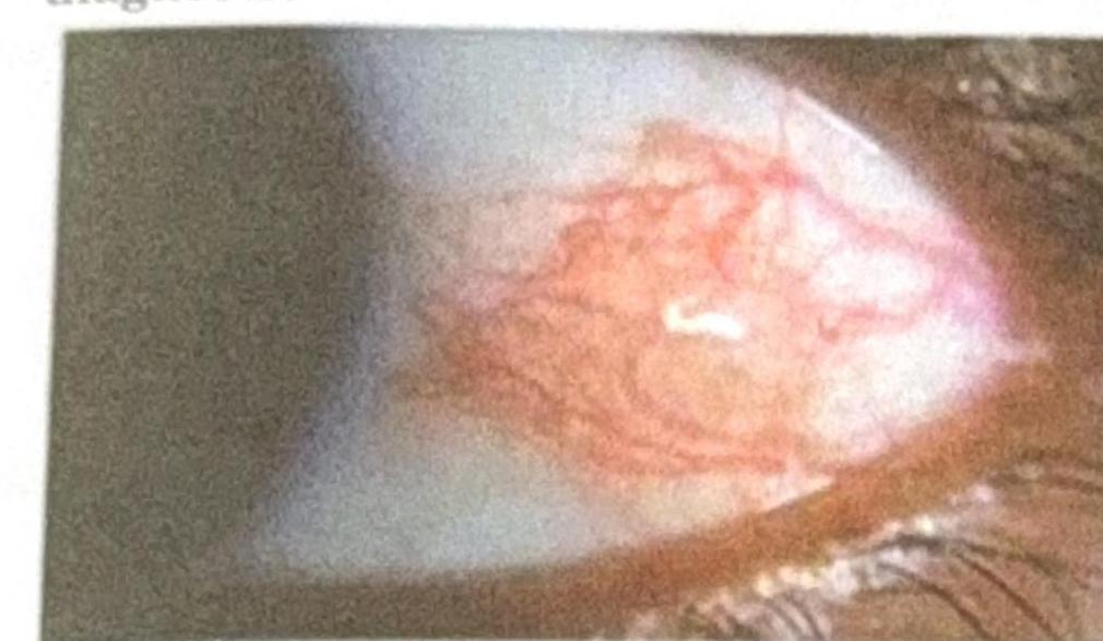
A 22-year-old Air-force test pilot presents after flying a sortie. He reports no pain or vision changes. Eye examination reveals a localized red patch on the sclera. What is the most likely diagnosis?
Blue sclera is seen in all of the following conditions except:
Evisceration is removal of which layer of eyeball?
Shortening of 2 mm of axial length of the eyeball causes?
Ciliary staphyloma is seen in which of the following conditions?
Explanation: Cartilage - **Type II collagen** is the predominant collagen found in hyaline and elastic cartilage (the typical forms of cartilage), providing their characteristic tensile strength and resilience [2]. - Type I collagen is NOT the primary collagen in cartilage, making this the correct answer. - Note: Fibrocartilage is a specialized form that does contain Type I collagen, but standard cartilage refers to hyaline and elastic types. *Ligament* - **Type I collagen** is the primary structural component of ligaments, providing high tensile strength to connect bones and stabilize joints. - Its presence allows ligaments to withstand significant pulling forces without stretching excessively. *Aponeurosis* - **Type I collagen** is abundant in aponeuroses, which are flat sheet-like tendons that connect muscles to bones or other muscles. - This type of collagen provides the necessary tensile strength for these broad connective tissues. *Bone* - **Type I collagen** is the most abundant collagen in bone matrix, accounting for approximately 90% of its organic content [1]. - It forms a robust scaffold that gives bone its flexibility and tensile strength, working in conjunction with mineralized components like hydroxyapatite [1].
Explanation: ***Keratin*** - **Keratin** is a fibrous, structural protein and is a prime example of a **scleroprotein** (also known as a **fibrous protein**). - It provides strength and structural integrity to tissues like hair, nails, and the outer layer of skin. *Zein* - **Zein** is a prolamine protein found in corn. - It is an **alcohol-soluble protein** and is not classified as a scleroprotein. *Glutenin* - **Glutenin** is a major storage protein found in wheat. - It is classified as a **globulin** and contributes to the elasticity of dough, but is not a scleroprotein. *Ovoglobulin* - **Ovoglobulin** refers to a group of globular proteins found in egg white. - These are typically **soluble in water** or dilute salt solutions and do not share the fibrous, structural characteristics of scleroproteins.
Explanation: ***10-21 mm Hg*** - This range is widely accepted as the **normal intraocular pressure (IOP)** in healthy individuals. - Maintaining IOP within this range is crucial for preventing damage to the **optic nerve** and conditions like **glaucoma**. *2.1-6 mm Hg* - This range is significantly **lower** than the normal physiological IOP. - Pressures in this range could indicate conditions like **hypotony**, which can lead to vision problems. *7-14 mm Hg* - While closer to the normal range, this range is still generally considered to be at the **lower end of normal** or slightly below. - Many individuals would fall within 10-21 mm Hg, making this a less accurate representation of the typical normal range. *16-32 mm Hg* - The upper part of this range (above 21 mm Hg) is considered **elevated IOP**, a significant risk factor for **glaucoma**. - Pressures above 21 mm Hg require closer monitoring and potentially treatment to prevent **optic nerve damage**.
Explanation: ***Osteogenesis imperfecta*** - **Osteogenesis imperfecta** (brittle bone disease) is the **MOST CLASSIC** condition associated with blue sclera. - Caused by a defect in **Type I collagen synthesis**, which is a major structural component of the sclera. - The deficiency in functional collagen leads to an **abnormally thin and translucent sclera**, allowing the underlying **choroidal pigment** to show through, giving it a characteristic blue appearance. - Blue sclera is seen in **80-90% of patients** with Type I osteogenesis imperfecta. *Buphthalmos* - **Buphthalmos** (congenital glaucoma) can occasionally show bluish sclera due to scleral thinning from increased intraocular pressure. - However, the primary findings are **corneal enlargement, corneal edema**, and increased eye size, not blue sclera as a defining feature. *Marfan's syndrome* - **Marfan's syndrome** is a connective tissue disorder caused by **fibrillin-1** defects. - Classic ocular features include **ectopia lentis** (lens dislocation), high myopia, and retinal detachment risk. - Blue sclera is **NOT a recognized clinical feature** of Marfan's syndrome. *Ehlers-Danlos syndrome* - This connective tissue disorder can present with blue sclera due to collagen abnormalities. - However, it is **less common** than osteogenesis imperfecta as a cause of blue sclera.
Explanation: ***Scleritis with rheumatoid arthritis*** - The image shows **deep, violaceous conjunctival injection** with surrounding edema and a nodular appearance near the limbus, consistent with **nodular scleritis**. Scleritis is inflammation of the sclera, often characterized by severe pain and association with systemic autoimmune diseases like **rheumatoid arthritis**, which can cause destructive lesions and recurrence. - The characteristic **deep vessel engorgement that does not blanch with phenylephrine** and the history of recurrence further support scleritis. *Episcleritis with rheumatoid arthritis* - Episcleritis presents with a **more superficial, bright red injection** involving the episclera, which generally **blanches with topical phenylephrine** and is less painful than scleritis. - While episcleritis can be associated with rheumatoid arthritis, the clinical features described (nodular swelling, deep injection) are more typical of scleritis. *Pinguecula* - A pinguecula is a **yellowish patch or bump** on the conjunctiva, typically on the nasal side of the eye, that is **not inflamed** unless irritated. - It consists of **degenerated collagen fibers** and elastic tissue, and does not present with the diffuse, deep vascular injection seen in the image. *Dry eye* - Dry eye is characterized by **ocular dryness, irritation, and sometimes a foreign body sensation**, but it typically causes **diffuse conjunctival hyperemia** rather than a localized, nodular, deep inflammation with surrounding edema as shown. - While dry eye can be associated with autoimmune diseases, its appearance is not consistent with the depicted nodular lesion.
Explanation: ***Subconjunctival hemorrhage*** - A **localized red patch on the sclera** with no pain or vision changes, especially after activities that can increase venous pressure (like flying a sortie or straining), is characteristic of a **subconjunctival hemorrhage**. - It results from the rupture of small blood vessels beneath the conjunctiva, causing blood to pool. *Hyphema* - This involves blood in the **anterior chamber of the eye**, usually visible as a fluid level and often causing pain or blurred vision. - It typically results from **trauma** and is not described as a localized red patch on the sclera. *Keratitis* - **Keratitis** is inflammation of the cornea, causing pain, redness, photophobia, and often blurred vision. - The patient has no pain or vision changes, and the presentation is a localized scleral patch, not diffuse corneal involvement. *Allergic conjunctivitis* - Presents with **redness**, itching, tearing, and often bilateral involvement, sometimes with discharge. - The description of a solitary, localized red patch without other allergic symptoms makes this diagnosis unlikely.
Explanation: ***Keratoconus*** - Keratoconus is a progressive eye disease in which the normally round cornea thins and begins to bulge into a cone-like shape, leading to **vision distortion**. - Blue sclera is **not a feature** of keratoconus. This is a **corneal condition** that does not affect the sclera. - Blue sclera, seen in the other conditions listed, occurs due to thinning of the sclera, making the underlying choroidal pigment visible. *Marfan's syndrome* - Patients with Marfan's syndrome can have blue sclera due to the **thinning of collagen** in the scleral tissue, allowing the underlying choroid to show through. - This connective tissue disorder affects multiple body systems, including the skeletal, cardiovascular, and ocular systems, with features like **arachnodactyly** and **aortic root dilation**. *Osteogenesis imperfecta* - Often referred to as **brittle bone disease**, osteogenesis imperfecta is characterized by defective **type I collagen synthesis**, which also affects the sclera. - The sclera becomes thin and translucent, revealing the underlying choroidal pigment, thus appearing **blue**. *Rheumatoid arthritis* - In rheumatoid arthritis, particularly with severe or long-standing disease, the sclera can become thinned due to **scleritis** or **scleromalacia perforans**. - This thinning can lead to a **blue discoloration** of the sclera, making the underlying choroid visible.
Explanation: ***Middle and inner*** - **Evisceration** involves the surgical removal of the internal contents of the eyeball, which include components derived from the **middle (uveal) and inner (retinal) layers**. - The **sclera** (outermost layer) and **extraocular muscles** are preserved, allowing for a more natural prosthetic eye fit. *All the layers of eyeball* - This describes **enucleation**, where the entire eyeball is removed including all three layers (sclera, choroid/ciliary body/iris, and retina). - Enucleation is a more extensive procedure than evisceration, typically performed for intraocular tumors or severe trauma where preservation of the sclera isn't possible. *Outer and inner* - This option is incorrect because the **outer layer (sclera)** is specifically preserved in evisceration. - Removing the outer layer would lead to a more destructive procedure, inconsistent with the definition of evisceration. *Outer and middle* - This option is incorrect because the **outer layer (sclera)** is preserved during evisceration, while the entire **inner layer (retina)** is removed. - The middle layer (uvea) is removed, but this option inaccurately states the fate of the outer and inner layers.
Explanation: ***6D hypermetropia*** - A 1 mm shortening of the **axial length** of the eyeball typically results in approximately **3 diopters** of hypermetropia. - Therefore, a 2 mm shortening would cause **6 diopters** (2 mm x 3 D/mm) of hypermetropia. *3D myopia* - Myopia (nearsightedness) is caused by an **eyeball that is too long** or a cornea that is too steeply curved, not by a shortened axial length. - A 2 mm shortening would cause **hypermetropia** (farsightedness), not myopia. *2D myopia* - This option incorrectly identifies both the **type of refractive error** (myopia instead of hypermetropia) and the magnitude of the change. - Shortening of the axial length makes the eye effectively **farsighted**, not nearsighted. *1D hypermetropia* - While reflecting the correct type of refractive error (hypermetropia), the **magnitude is incorrect**. - A 1 mm change in axial length results in about 3 diopters, so 2 mm would be **6 diopters**, not 1 diopter.
Explanation: **Explanation:** **Ciliary staphyloma** is a localized thinning and bulging of the sclera lined by the underlying ciliary body. It occurs due to a combination of weakened scleral integrity and chronically elevated intraocular pressure (IOP). 1. **Why Absolute Glaucoma is correct:** In absolute glaucoma, the IOP is severely and chronically elevated. This persistent pressure causes the sclera to stretch and thin, particularly in the **ciliary zone** (the area 2–8 mm behind the limbus). As the sclera thins, the dark pigment of the underlying ciliary body shines through, giving it a characteristic bluish-black appearance. 2. **Why the other options are incorrect:** * **Pathological Myopia:** This is typically associated with **Posterior staphyloma**, where the thinning occurs at the posterior pole (macular area) due to excessive axial elongation. * **Retinoblastoma:** While advanced tumors can cause globe enlargement (buphthalmos) or extraocular extension, they do not typically present as a focal ciliary staphyloma. * **Episcleritis:** This is a self-limiting, superficial inflammation of the episcleral tissues that does not lead to scleral thinning or staphyloma formation. **Clinical Pearls for NEET-PG:** * **Types of Staphyloma:** * **Anterior (Intercalary):** At the limbus; seen in secondary glaucoma following corneal perforation. * **Ciliary:** 2–8 mm from limbus; seen in Absolute Glaucoma and Scleritis. * **Equatorial:** At the exit of vortex veins; seen in Pathological Myopia. * **Posterior:** At the posterior pole; hallmark of Pathological Myopia. * **Key Sign:** The "bluish" color of a staphyloma is not due to the sclera itself, but the **uveal tissue** visible through the thinned sclera.
Get full access to all questions, explanations, and performance tracking.
Start For Free