
Diseases of the Retina — MCQs

On this page
Which of the following is an ocular emergency?
A prematurely born baby at 29 weeks is examined at 42 weeks post-conceptional age and shows stage 2 zone 1 ROP with plus disease in both eyes. How will you manage this patient?
Which of the following statements is FALSE regarding Stargardt's disease?
A myopic patient presents with complaints of flashes and floaters. On examination, a deep anterior chamber is seen. What is the likely diagnosis?
A patient presents with vision problems and has a history of cataract surgery. OCT finding is shown below. What is the syndrome most likely associated with these findings?
Fundoscopy findings are shown in the image below. What is the most likely diagnosis?
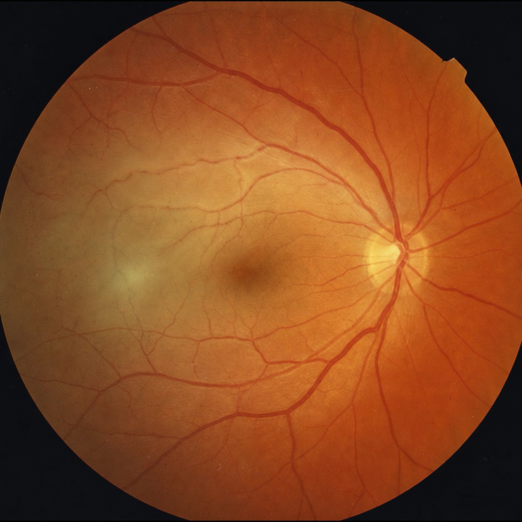
Fundoscopy findings are shown in the image below. What is the most likely diagnosis? 
A 62-year-old diabetic patient presents with sudden painless loss of vision in the right eye. Fundoscopy reveals a pale, edematous retina with a cherry-red spot at the macula. Retinal arterioles appear narrowed. Visual acuity is light perception only. What is the most likely diagnosis?
Which of the following is seen in proliferative diabetic retinopathy?
Which of the following investigations allows examination of all layers of the retina?
Practice by Chapter
Retinal Anatomy and Physiology
Practice Questions
Age-Related Macular Degeneration
Practice Questions
Diabetic Retinopathy
Practice Questions
Retinal Vascular Diseases
Practice Questions
Retinal Detachment
Practice Questions
Hereditary Retinal Dystrophies
Practice Questions
Inflammatory Retinal Diseases
Practice Questions
Retinal Tumors
Practice Questions
Retinopathy of Prematurity
Practice Questions
Retinal Imaging Techniques
Practice Questions
Intravitreal Pharmacotherapy
Practice Questions
Vitreoretinal Surgery
Practice Questions
Want unlimited practice?
Get full access to all questions, explanations, and performance tracking.
Scan to download app