Which of the following conditions is associated with Macular edema?
Regarding Retinitis pigmentosa, which of the following statements is FALSE?
What is the most common condition of inherited blindness due to a mitochondrial chromosomal anomaly?
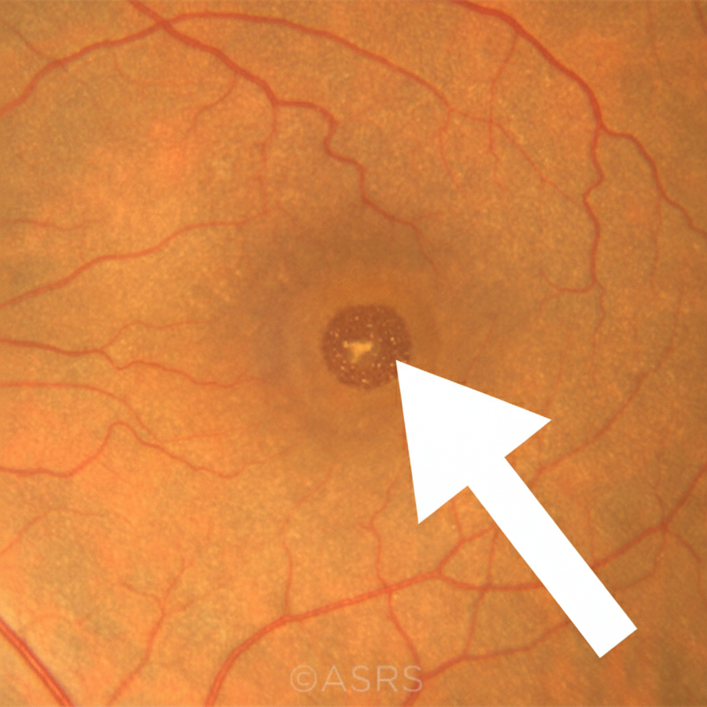
What finding in a fundus examination of a myopic patient is indicated by an arrow?

What is the cause of blindness in Central Retinal Vein Occlusion (CRVO)?
Malignant change in a choroidal nevus is evidenced by:
All of the following statements are true regarding indirect ophthalmoscopy, except:
Which of the following is not a complication of Eale's disease?
Ocriplasmin is the newer drug used in which of the following conditions?
Which of the following ocular conditions is characterized by calcification as a pathognomonic feature?
Explanation: **Explanation:** **Macular Edema** occurs due to the breakdown of the blood-retinal barrier, leading to the accumulation of fluid in the intraretinal layers of the macula. **Why Diabetes Mellitus is Correct:** Diabetic Macular Edema (DME) is the most common cause of vision loss in patients with Diabetic Retinopathy. Chronic hyperglycemia leads to pericyte loss and endothelial damage, causing capillary leakage. This results in fluid accumulation within the Henle’s layer (outer plexiform layer), often presenting as **Cystoid Macular Edema (CME)**. **Analysis of Incorrect Options:** * **A. Central Retinal Artery Occlusion (CRAO):** This presents with **cloudy swelling** (intracellular edema) of the entire retina due to ischemia, not localized macular edema. The classic finding is a "Cherry Red Spot" because the fovea is thin and reflects the underlying vascular choroid. * **B. Papilledema:** This refers specifically to bilateral swelling of the **optic disc** due to increased intracranial pressure. While severe cases (Grade IV/V) may show macular fan/star formation (exudates), the primary pathology is disc edema, not macular edema. * **D. Age-related Macular Degeneration (ARMD):** Dry ARMD is characterized by **Drusen** and geographic atrophy. While Wet (Neovascular) ARMD involves subretinal fluid/hemorrhage due to CNVM, "Macular Edema" as a standalone clinical entity is most classically associated with systemic vascular diseases like Diabetes. **High-Yield Clinical Pearls for NEET-PG:** * **Gold Standard Investigation:** Optical Coherence Tomography (OCT) is used to quantify macular thickness. * **First-line Treatment for DME:** Intravitreal Anti-VEGF injections (e.g., Ranibizumab, Aflibercept). * **Irvine-Gass Syndrome:** A specific type of CME occurring after cataract surgery. * **Histological Site:** Fluid in CME typically accumulates in the **Outer Plexiform Layer (Henle’s layer)**.
Explanation: **Explanation:** Retinitis Pigmentosa (RP) is a hereditary, progressive dystrophy primarily affecting the **rod photoreceptors** first, followed by the cones. **Why Option C is the correct (False) statement:** In RP, the degeneration starts in the mid-peripheral retina where rod density is highest. Consequently, the initial symptoms are **nyctalopia (night blindness)** and **peripheral field loss** (ring scotoma). Central vision is typically **preserved** until the late stages of the disease when cone involvement becomes significant or complications like Cystoid Macular Edema (CME) occur. Therefore, early loss of central vision is not a common initial symptom. **Analysis of other options:** * **Option A:** Night blindness is the hallmark initial symptom due to early rod dysfunction. * **Option B & D:** These describe the classic **"Diagnostic Triad"** of RP fundus findings: 1. **Bony spicule pigmentation** (perivascular pigment clumps in the mid-periphery). 2. **Arteriolar attenuation** (narrowing of retinal vessels). 3. **Waxy pallor of the optic disc** (due to consecutive optic atrophy). **NEET-PG High-Yield Pearls:** * **Inheritance:** Most common is Autosomal Recessive; most severe is X-linked. * **Visual Field:** Characteristically shows a **Ring Scotoma**. * **ERG Findings:** The Electroretinogram (ERG) is **subnormal or extinguished** even before fundus changes appear (earliest diagnostic tool). * **Associated Syndromes:** **Usher Syndrome** (RP + Sensorineural hearing loss) and **Laurence-Moon-Biedl Syndrome** (RP + Obesity + Polydactyly + Hypogonadism + Mental retardation). * **Ocular Associations:** Posterior subcapsular cataract, Myopia, and Open-angle glaucoma.
Explanation: ### Explanation **Correct Answer: D. Leber’s Hereditary Optic Neuropathy (LHON)** **Why it is correct:** Leber’s Hereditary Optic Neuropathy (LHON) is the most common cause of inherited blindness resulting from a **mitochondrial DNA (mtDNA) mutation**. It typically involves point mutations at positions 11778, 3460, or 14484. Because mitochondria are inherited exclusively from the oocyte, LHON follows a **maternal inheritance pattern** (all children of an affected mother are at risk, but affected fathers do not pass it on). It clinically presents as painless, subacute, bilateral central vision loss, primarily affecting young males. **Why the other options are incorrect:** * **A. Retinopathy of Prematurity (ROP):** This is an acquired proliferative retinopathy caused by oxygen toxicity and prematurity; it is not a genetic or mitochondrial disorder. * **B. Retinitis Pigmentosa (RP):** While RP is a common cause of inherited blindness, it is primarily caused by mutations in **nuclear DNA** (Autosomal Dominant, Recessive, or X-linked), not mitochondrial DNA. * **C. Retinal Detachment:** This is a structural/mechanical emergency often related to trauma, high myopia, or vitreous changes, rather than a chromosomal anomaly. **High-Yield Clinical Pearls for NEET-PG:** * **Inheritance:** Maternal inheritance (Mitochondrial). * **Demographics:** Strong male predilection (approx. 80-90%), though the mutation is carried by females. * **Fundus Findings:** Circumpapillary telangiectatic microangiopathy and swelling of the nerve fiber layer (pseudo-edema), which does not leak on Fluorescein Angiography (FFA). * **Key Association:** Often associated with "Leber’s plus" syndromes, which may include cardiac conduction defects (arrhythmias) and neurological abnormalities.
Explanation: ***Foster-Fuchs spot*** - A **dark pigmented macular lesion** seen in high myopia, resulting from **subretinal neovascularization** followed by hemorrhage and subsequent **retinal pigment epithelium (RPE) hyperplasia**. - Appears as a **well-defined dark spot** in the macular area on fundus examination, representing the end-stage of **choroidal neovascular membrane** formation in pathologic myopia. *Lacquer cracks* - These are **linear breaks in Bruch's membrane** that appear as yellowish or whitish streaks, not dark pigmented spots. - They represent **mechanical breaks** due to excessive axial elongation in high myopia, appearing as **crack-like linear lesions** rather than focal dark spots. *Arlt line* - This is a **conjunctival scar** associated with **trachoma**, not a fundus finding at all. - It appears as a **horizontal line of scarring** in the superior tarsal conjunctiva, completely unrelated to myopic retinal changes. *Myopic crescent* - This refers to **peripapillary atrophy** around the optic disc, appearing as a pale crescent-shaped area. - It represents **chorioretinal atrophy** adjacent to the optic nerve head, not a macular pigmented lesion as indicated by the arrow.
Explanation: **Explanation:** Central Retinal Vein Occlusion (CRVO) is a common retinal vascular disorder characterized by the "blood and thunder" fundus appearance. Blindness or severe visual loss in CRVO is multifactorial, resulting from both acute changes and long-term complications. **Why "All of the above" is correct:** 1. **Cystoid Macular Edema (CME):** This is the **most common cause** of decreased vision in both ischemic and non-ischemic CRVO. Increased intraluminal pressure leads to the leakage of fluid into the Henle’s layer of the macula, causing structural damage and loss of central acuity. 2. **Macular Hemorrhage:** In the acute phase, extensive deep and superficial (flame-shaped) hemorrhages can involve the fovea, directly blocking photoreceptors and leading to a sudden drop in vision. 3. **Vitreous Hemorrhage:** This is a late-stage complication primarily seen in **Ischemic CRVO**. Retinal ischemia triggers the release of Vascular Endothelial Growth Factor (VEGF), leading to Neovascularization of the Disc (NVD) or elsewhere (NVE). These fragile new vessels can rupture, causing vitreous hemorrhage and sudden vision loss. **Clinical Pearls for NEET-PG:** * **The "90-Day Glaucoma":** Ischemic CRVO is highly associated with Neovascular Glaucoma (NVG), which typically develops around 3 months after the occlusion. * **Investigation of Choice:** **Optical Coherence Tomography (OCT)** is used to monitor Macular Edema, while **Fundus Fluorescein Angiography (FFA)** is the gold standard to differentiate between Ischemic and Non-ischemic types (based on areas of capillary non-perfusion). * **Management:** Intravitreal Anti-VEGF injections (e.g., Ranibizumab, Aflibercept) are the mainstay of treatment for macular edema. Pan-retinal photocoagulation (PRP) is indicated if neovascularization develops.
Explanation: **Explanation:** The transformation of a benign choroidal nevus into a **malignant melanoma** is a critical clinical progression. To differentiate between a stable nevus and an early melanoma, clinicians use the **"TFSOM" mnemonic** (To Find Small Ocular Melanoma). * **Increased height and pigmentation (Option A):** A thickness of >2 mm is a major risk factor. Rapid growth or an increase in the density of pigmentation suggests active cellular proliferation characteristic of malignancy. * **Orange pigment (Option B):** This represents the accumulation of **lipofuscin** at the level of the Retinal Pigment Epithelium (RPE). It indicates metabolic distress and chronic RPE damage caused by an underlying growing tumor. This is one of the most predictive signs of malignancy. * **Serous detachment (Option C):** Malignant cells disrupt the overlying RPE and the blood-retinal barrier, leading to the leakage of subretinal fluid. The presence of subretinal fluid (serous detachment) is a hallmark of activity and transformation. Since all three features are established clinical indicators of malignant transformation, **Option D** is the correct answer. **High-Yield Clinical Pearls (TFSOM Mnemonic):** To remember the risk factors for malignant change, use: **"To Find Small Ocular Melanoma Using Helpful Hints Daily"** * **T** – **T**hickness >2 mm * **F** – Subretinal **F**luid * **S** – **S**ymptoms (flashes/floaters/blurred vision) * **O** – **O**range pigment (Lipofuscin) * **M** – **M**argin within 3 mm of the optic disc * **UH** – **U**ltrasonographic **H**ollow (low internal reflectivity) * **D** – Absence of **D**rusen (Drusen usually indicate a long-standing, stable, benign nevus).
Explanation: ### Explanation In Indirect Ophthalmoscopy, a strong light source and a convex condensing lens (usually +20D) are used to create a **real, inverted, and magnified image** of the fundus between the lens and the examiner. **Why Option C is the "Except" (Correct Answer):** The question contains a technical error in its framing or the provided key. In clinical practice, indirect ophthalmoscopy is **superior** to direct ophthalmoscopy for hazy media because of its high-intensity illumination. However, if we analyze the options based on standard textbook definitions: * **Option B (There is no stereopsis)** is actually a **False** statement. Indirect ophthalmoscopy provides excellent **stereopsis** (3D visualization) because it is a binocular procedure. * **Option D (The retinal periphery cannot be examined)** is also a **False** statement. Indirect ophthalmoscopy is the gold standard for examining the retinal periphery up to the **ora serrata** (especially with scleral indentation). *Note: In many competitive exams, if the question asks for the "False" statement and lists "No stereopsis" or "Periphery cannot be examined," those are the intended answers. If Option C is marked correct in your key, it implies a pedagogical error, as indirect ophthalmoscopy is specifically indicated for hazy media.* **Analysis of Other Options:** * **Option A:** True. It can be performed while the patient is sitting, reclining, or supine. * **Option B:** False. It provides a binocular view, hence **stereopsis is present**. * **Option D:** False. It is the preferred method for **peripheral retinal examination**. **NEET-PG High-Yield Pearls:** 1. **Image Characteristics:** Real, Inverted (both vertically and laterally), and formed between the lens and the examiner. 2. **Magnification:** Approximately 3x with a +20D lens (Magnification = 60 / Power of lens). 3. **Field of View:** Large (approx. 8 disc diameters), making it ideal for a general overview. 4. **Condensing Lenses:** +20D is standard; +13D gives more magnification; +28D/30D gives a wider field of view.
Explanation: **Explanation:** **Eales’ Disease** is an idiopathic, inflammatory peripheral perivasculitis (primarily affecting the venules) that typically affects young healthy males. The pathophysiology involves peripheral retinal ischemia, which leads to the release of Vasoproliferative factors (VEGF). **Why Optic Neuritis is the correct answer:** Optic neuritis is an inflammatory condition of the optic nerve, often associated with multiple sclerosis or infections. It is **not** a feature of Eales’ disease. While Eales’ disease involves inflammation, it is localized to the retinal vasculature (periphlebitis) and does not primarily involve the optic nerve fibers. **Why the other options are incorrect:** The complications of Eales’ disease follow a sequence driven by **retinal ischemia**: * **Retinal Detachment (B):** Recurrent vitreous hemorrhages lead to the formation of fibrovascular tractional bands. These bands can cause **Tractional Retinal Detachment**, a common late-stage complication. * **Rubeosis Iridis (A) & Neovascular Glaucoma (C):** Severe, chronic peripheral ischemia triggers neovascularization not just in the retina, but also on the iris (Rubeosis iridis). These vessels can block the aqueous outflow in the anterior chamber angle, resulting in **Neovascular Glaucoma (NVG)**. **Clinical Pearls for NEET-PG:** * **Demographics:** Most common in males aged 20–40 years. * **Stages:** Perivasculitis (sheathing) → Non-perfusion → Neovascularization → Complications (Vitreous hemorrhage/RD). * **Classic Sign:** "Sea-fan" neovascularization (similar to Sickle Cell Retinopathy). * **Association:** Historically linked to **Tuberculosis** sensitivity (positive Mantoux test), though the exact etiology remains idiopathic. * **Management:** Photocoagulation (LASER) for ischemia and Anti-VEGF for neovascularization.
Explanation: **Explanation:** **Ocriplasmin** is a recombinant truncated form of human **plasmin**. It is the first pharmacological agent approved for the treatment of symptomatic **Vitreomacular Traction (VMT)** and small macular holes. **Why Option B is Correct:** The underlying mechanism of VMT involves an incomplete posterior vitreous detachment (PVD) where the vitreous remains abnormally adherent to the macula, causing mechanical distortion. Ocriplasmin works through **enzymatic vitreolysis**. It targets and breaks down protein components (laminin, fibronectin, and collagen) at the vitreoretinal interface. By dissolving these "glue-like" proteins, it facilitates the release of traction and promotes a complete PVD without the need for surgical vitrectomy. **Why Other Options are Incorrect:** * **A. Retinal break:** These are treated with laser photocoagulation or cryopexy to create a chorioretinal scar. Ocriplasmin could theoretically worsen a break by inducing vitreous movement. * **C & D. Submacular/Diabetic macular bleed:** These conditions involve vascular pathology or neovascularization. Management typically involves Anti-VEGF agents (like Ranibizumab or Aflibercept) or surgical evacuation (for submacular hemorrhage), not enzymatic vitreolysis. **High-Yield Clinical Pearls for NEET-PG:** * **Route of Administration:** Intravitreal injection (0.125 mg). * **Indications:** Best results are seen in patients with focal VMT (adhesion ≤1500 µm) and those without an epiretinal membrane (ERM). * **Side Effects:** Patients may report "chromatopsia" (yellowish vision) or transient electroretinogram (ERG) changes post-injection. * **Alternative Treatment:** Pars Plana Vitrectomy (PPV) remains the gold standard for severe or non-responsive VMT.
Explanation: ### Explanation **Correct Answer: C. Retinoblastoma** **Why it is correct:** Retinoblastoma (RB) is the most common primary intraocular malignancy of childhood. **Calcification** is considered a pathognomonic hallmark of this tumor, occurring in approximately **90-95%** of cases. It results from DNA-calcium binding within areas of tumor necrosis (dystrophic calcification). On clinical examination, these appear as "chalky white" deposits. In NEET-PG, the presence of intraocular calcification in a child under 3 years old on a CT scan or B-scan ultrasound is virtually diagnostic of Retinoblastoma. **Why the other options are incorrect:** * **A. Malignant Melanoma of the Choroid:** This is the most common primary intraocular tumor in adults. Unlike RB, it rarely shows calcification. On imaging, it typically presents as a mushroom or collar-stud shaped mass with low internal reflectivity on USG. * **B. Retinocytoma:** This is a benign version (retinoma) of the same genetic mutation (RB1 gene). While it can show some calcification and "fish-flesh" appearance, it is a rare, non-progressive lesion and is not the classic answer for pathognomonic calcification in a malignant context. * **D. Angiomatosis Retinae (von Hippel-Lindau disease):** This is characterized by retinal hemangioblastomas (vascular tumors) with feeder vessels and exudation, but calcification is not a primary feature. **High-Yield Clinical Pearls for NEET-PG:** * **Most common sign:** Leukocoria (White pupillary reflex). * **Second most common sign:** Strabismus. * **Histology:** Look for **Flexner-Wintersteiner rosettes** (highly specific) and Homer-Wright rosettes. * **Imaging Choice:** **B-scan Ultrasonography** is the initial screening tool; **CT scan** is excellent for detecting calcification; **MRI** is preferred to evaluate optic nerve involvement and pinealoblastoma (Trilateral Retinoblastoma). * **Inheritance:** 40% are germline (bilateral/multifocal), 60% are somatic (unilateral).
Retinal Anatomy and Physiology
Practice Questions
Age-Related Macular Degeneration
Practice Questions
Diabetic Retinopathy
Practice Questions
Retinal Vascular Diseases
Practice Questions
Retinal Detachment
Practice Questions
Hereditary Retinal Dystrophies
Practice Questions
Inflammatory Retinal Diseases
Practice Questions
Retinal Tumors
Practice Questions
Retinopathy of Prematurity
Practice Questions
Retinal Imaging Techniques
Practice Questions
Intravitreal Pharmacotherapy
Practice Questions
Vitreoretinal Surgery
Practice Questions
Get full access to all questions, explanations, and performance tracking.
Start For Free