
Diseases of the Retina — MCQs

On this page
Amster grid is used in which type of examination?
Epiretinal membrane is seen in which of the following conditions?
Which of the following is NOT a cause of night blindness?
The "shower of golden rain" appearance in synchisis scintillans is due to which of the following?
What is the cause of acute vision loss in a patient with alcoholic pancreatitis?
Black floaters in a diabetic patient indicate what condition?
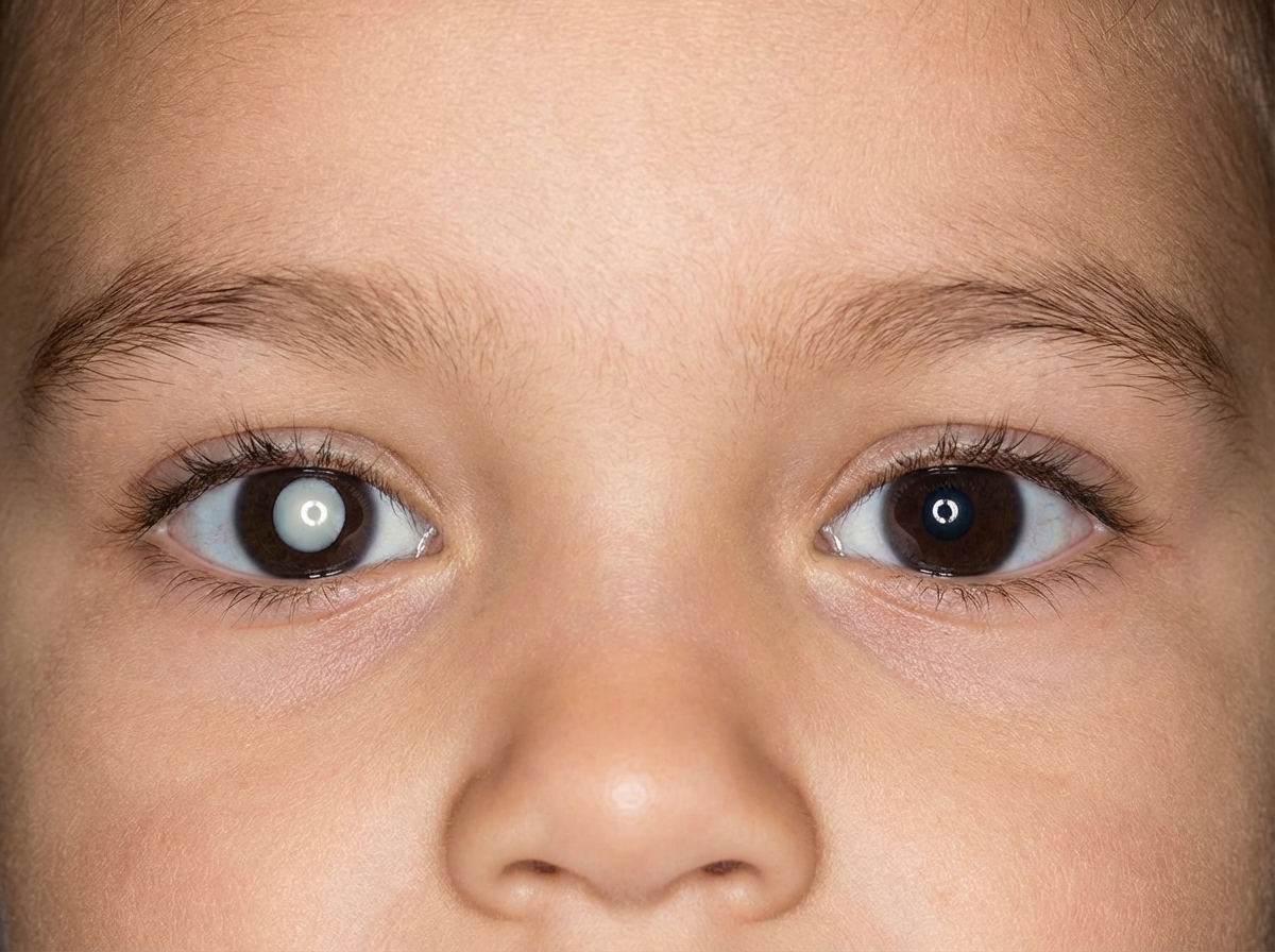
A 2-year-old child presented with a specific abnormality, and there is a history of a similar illness in his father. What is the most likely underlying condition responsible?
Lattice degeneration is seen in which of the following conditions?
All of the following are features of asteroid hyalosis except?
Vitreous hemorrhage produces which of the following?
Practice by Chapter
Retinal Anatomy and Physiology
Practice Questions
Age-Related Macular Degeneration
Practice Questions
Diabetic Retinopathy
Practice Questions
Retinal Vascular Diseases
Practice Questions
Retinal Detachment
Practice Questions
Hereditary Retinal Dystrophies
Practice Questions
Inflammatory Retinal Diseases
Practice Questions
Retinal Tumors
Practice Questions
Retinopathy of Prematurity
Practice Questions
Retinal Imaging Techniques
Practice Questions
Intravitreal Pharmacotherapy
Practice Questions
Vitreoretinal Surgery
Practice Questions
Want unlimited practice?
Get full access to all questions, explanations, and performance tracking.
Scan to download app