
Diseases of the Retina — MCQs

On this page
Angioid streaks are seen in which of the following conditions?
Schaffer's sign is seen in which condition?
Which of the following is NOT a sign of diabetic retinopathy?
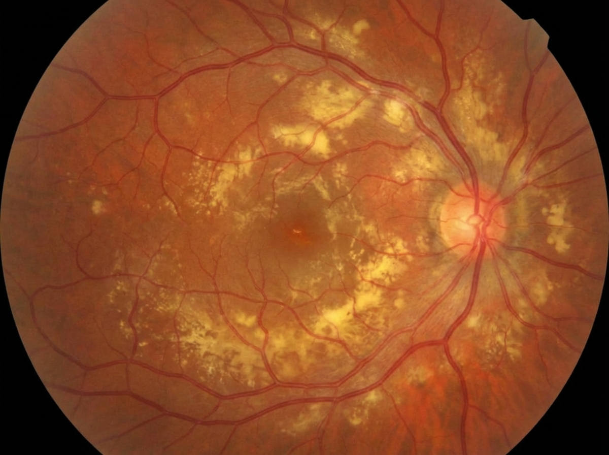
A 3-year-old male presents with sudden onset of visual loss in the left eye. Ophthalmological examination reveals strabismus and a white pupillary reflex. Fundus findings are as shown below. What is your diagnosis?
Retinitis pigmentosa inheritance is by which mode?
Regarding retinoblastoma, all of the following are true, EXCEPT?
Which of the following is associated with Retinoblastoma?
Which of the following is NOT a prognostic factor in choroidal melanoma?
Which of the following is NOT a cause of exudative retinal detachment?
Sudden loss of vision in a patient with diabetic retinopathy is most commonly due to what condition?
Practice by Chapter
Retinal Anatomy and Physiology
Practice Questions
Age-Related Macular Degeneration
Practice Questions
Diabetic Retinopathy
Practice Questions
Retinal Vascular Diseases
Practice Questions
Retinal Detachment
Practice Questions
Hereditary Retinal Dystrophies
Practice Questions
Inflammatory Retinal Diseases
Practice Questions
Retinal Tumors
Practice Questions
Retinopathy of Prematurity
Practice Questions
Retinal Imaging Techniques
Practice Questions
Intravitreal Pharmacotherapy
Practice Questions
Vitreoretinal Surgery
Practice Questions
Want unlimited practice?
Get full access to all questions, explanations, and performance tracking.
Scan to download app