In diabetic retinopathy, which layer of the retina is primarily affected?
Assertion: Vitamin D analogues are effective in psoriasis. Reason: They reduce keratinocyte proliferation
A patient presents with a central scotoma and metamorphopsia. What is the most likely cause?
Most common age related change in vitreous?
Fluorescein dye for ophthalmological diagnosis is injected into:
SAFE strategy is recommended for-
What type of deposit is commonly associated with age-related macular degeneration?
In the context of ophthalmology, the Amsler grid is primarily used for:
Ranibizumab is a monoclonal antibody against?
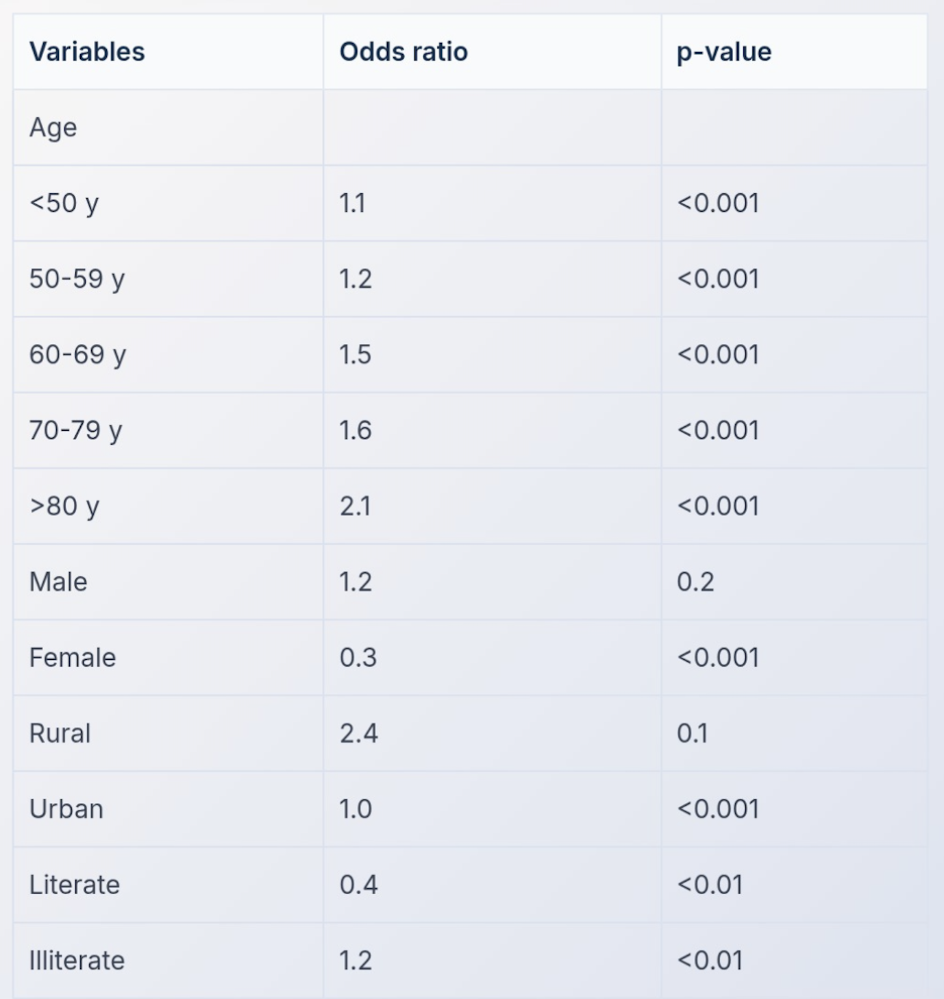
A multivariate analysis was conducted to examine the relationship between risk of developing blindness and age. The results are shown in the table below. Which of the following is true?

Explanation: ***Inner nuclear layer*** - The inner nuclear layer contains the **retinal capillary network**, which is the primary site of pathology in diabetic retinopathy. - **Microangiopathy** (pericyte loss, basement membrane thickening, endothelial cell damage) occurs in the capillaries located within this layer. - **Microaneurysms**, the earliest ophthalmoscopic sign of diabetic retinopathy, form from damaged capillaries in the inner nuclear layer. - **Diabetic macular edema (DME)** involves fluid accumulation that begins at the level of the capillaries in the inner nuclear and inner plexiform layers, then extends to the outer plexiform layer. *Outer plexiform layer* - This layer is **secondarily affected** by leakage from damaged capillaries in deeper retinal layers (inner nuclear and inner plexiform layers). - **Hard exudates** (lipid and protein deposits) accumulate in the outer plexiform layer as a consequence of capillary leakage, but this is not the primary site of vascular pathology. - The outer plexiform layer itself has minimal vasculature and is not where the initial microvascular changes occur. *Layer of rods and cones* - Photoreceptors are affected only in advanced stages of diabetic retinopathy due to chronic ischemia and secondary damage. - The primary pathology is vascular and occurs in the inner retinal layers where capillaries are located, not in the avascular photoreceptor layer. *Retinal pigment epithelium* - The RPE is not directly affected by the microvascular changes that characterize diabetic retinopathy. - RPE dysfunction is more characteristic of **age-related macular degeneration (AMD)** and other degenerative conditions. - In diabetic retinopathy, the RPE may be affected indirectly in very advanced cases but is not a primary site of pathology.
Explanation: ***Both A & R true, R explains A*** - **Vitamin D analogues** (e.g., calcipotriol) are a cornerstone treatment for psoriasis because they effectively modulate **keratinocyte proliferation** and differentiation. - Psoriasis is characterized by the **rapid overgrowth of keratinocytes**, and the antiproliferative effects of vitamin D analogues directly address this pathological hallmark. *A false R true* - This option is incorrect because both the assertion (Vitamin D analogues are effective in psoriasis) and the reason (They reduce keratinocyte proliferation) are individually true. - The effectiveness of vitamin D analogues in treating psoriasis is well-established in dermatological practice. *Both A & R true, R doesn't explain A* - This option is incorrect because the reduction of keratinocyte proliferation is precisely *how* vitamin D analogues exert their therapeutic effect in psoriasis. - The mechanism of action described in the reason directly explains the efficacy mentioned in the assertion. *A true R false* - This option is incorrect because the reason ("They reduce keratinocyte proliferation") is a fundamental and well-understood mechanism by which vitamin D analogues work in psoriasis. - Vitamin D analogues bind to vitamin D receptors in keratinocytes, influencing gene expression to inhibit their excessive growth.
Explanation: ***Macular degeneration*** - **Central scotoma** and **metamorphopsia** are classic symptoms of macular degeneration, indicating damage to the macula which is responsible for central and detailed vision. - **Metamorphopsia** refers to distorted vision, where straight lines appear wavy, and is a key indicator of macular involvement. *Retinal detachment* - While a significant eye condition, **retinal detachment** typically presents with symptoms like a sudden increase in **floaters**, flashes of light (photopsia), and a "curtain" coming across the field of vision, not primarily central scotoma and metamorphopsia. - It involves the separation of the sensory retina from the underlying retinal pigment epithelium, causing peripheral visual field loss initially, which can progress to central loss if the macula becomes detached. *Glaucoma* - **Glaucoma** is characterized by progressive optic nerve damage, often associated with increased intraocular pressure. - It typically leads to **peripheral vision loss** first, often described as tunnel vision, and usually does not cause metamorphopsia or a central scotoma until very late stages. *Optic neuritis* - **Optic neuritis** involves inflammation of the optic nerve, leading to sudden, often painful, vision loss in one eye and a **central scotoma**. - However, it typically does not cause **metamorphopsia**, which is more specific to macular pathology.
Explanation: ***Posterior vitreous detachment (PVD)*** - As we age, the **vitreous body** undergoes liquefaction and shrinkage, leading to its separation from the **retina**, most commonly posteriorly. - This is a very common and usually benign age-related phenomenon, often presenting with **floaters** and **flashes of light**. *Anterior vitreous detachment* - **Anterior vitreous detachment** is much less common than PVD and not typically considered the most frequent age-related change. - It often occurs secondary to **trauma** or specific surgical procedures rather than spontaneous aging alone. *Vitreous hemorrhage* - **Vitreous hemorrhage** is a pathological condition involving bleeding into the vitreous humor, often due to **diabetic retinopathy**, retinal tears, or trauma. - It is not a normal age-related change but a complication of underlying disease. *Vitritis* - **Vitritis** refers to inflammation of the vitreous humor, commonly associated with **uveitis** or infections. - It is an inflammatory disease process, not a physiological age-related degeneration of the vitreous.
Explanation: ***Antecubital vein*** - Fluorescein angiography requires rapid delivery of the dye into the **systemic circulation** for quick visualization of retinal and choroidal vasculature. - The **antecubital vein** is a readily accessible, large superficial vein that allows for quick and efficient intravenous dye injection. *Popliteal vein* - The popliteal vein is located behind the **knee** and is not a standard or practical site for routine intravenous injections, especially when rapid delivery to the eye is needed. - Its location makes it less accessible and potentially more uncomfortable for the patient compared to an arm vein. *Femoral vein* - The femoral vein is a large, deep vein in the **groin**, typically reserved for central venous access or specific procedures due to the increased risk of complications like infection or hematoma. - It is not routinely used for peripheral intravenous injections such as fluorescein, where a more superficial and accessible vein is preferred. *Subclavian vein* - The subclavian vein is a **central vein** located under the clavicle, accessed via a more invasive procedure, usually for central venous catheters or hemodialysis access. - It carries higher risks compared to peripheral venipuncture and is not chosen for simple diagnostic dye injections like fluorescein.
Explanation: ***Trachoma*** * The **SAFE strategy (Surgery, Antibiotics, Facial Cleanliness, Environmental improvement)** is the WHO-recommended public health approach for the elimination of **trachoma**, a chronic eye infection caused by *Chlamydia trachomatis*. * This comprehensive strategy addresses both active infection and its blinding sequelae, specifically **trichiasis** (in-turned eyelashes) through surgery. *Diabetic retinopathy* * Management of diabetic retinopathy primarily involves **blood sugar control, regular ophthalmologic exams, laser photocoagulation, and anti-VEGF injections**, not the SAFE strategy. * The focus is on preventing and treating retinal damage caused by **diabetes**, which is distinct from infectious causes. *Glaucoma* * Glaucoma is characterized by **optic nerve damage** and visual field loss, usually due to elevated intraocular pressure, and is managed with **medication, laser therapy, or surgery (e.g., trabeculectomy)**. * It is a **neurodegenerative condition**, not an infectious disease, so the SAFE strategy is not applicable. *Cataract* * Cataracts involve the **clouding of the natural lens** of the eye, leading to blurred vision, and are primarily treated through **surgical removal of the cloudy lens** and implantation of an artificial intraocular lens. * This condition is age-related or can be caused by trauma or disease, but it is **not an infection** for which the SAFE strategy would be relevant.
Explanation: ***Drusen*** - **Drusen** are yellow deposits of extracellular material that accumulate beneath the **retinal pigment epithelium (RPE)**. - Their presence is a hallmark sign of **age-related macular degeneration (AMD)** and can lead to vision loss by disrupting retinal function. *Iron* - While iron can accumulate in ocular tissues in conditions like **siderosis bulbi** (due to retained intraocular foreign bodies), it is not a characteristic deposit of macular degeneration. - Ocular iron deposition typically causes different pathologies, such as retinal dysfunction or glaucoma, rather than AMD. *Lipochrome* - **Lipochrome** refers to a class of pigments, including **lipofuscin**, which can accumulate in cells as a byproduct of cellular metabolism and aging. - Although lipofuscin buildup occurs in the RPE with age, **drusen** are the specific, organized extracellular deposits pathognomonic for macular degeneration. *Hemosiderine* - **Hemosiderin** is an iron-storage complex formed from the breakdown of hemoglobin, found in situations of hemorrhage or chronic bleeding. - It is not a typical deposit found in macular degeneration; its presence in the retina usually indicates a history of retinal hemorrhage.
Explanation: ***Identifying central vision defects*** - The Amsler grid is specifically designed to detect **distortions** or **scotomas** (blind spots) in the **central visual field**, which is crucial for tasks like reading and recognizing faces. - It is frequently used for monitoring conditions affecting the macula, such as **age-related macular degeneration (AMD)**, where patients might perceive straight lines as wavy or missing. *Evaluating optic disc morphology* - **Optic disc morphology** is typically assessed with an **ophthalmoscope** or by imaging techniques like **optical coherence tomography (OCT)**, which provide detailed views of the optic nerve head. - The Amsler grid does not provide direct visualization or measurement of the optic disc's structure. *Assessing eye alignment issues* - Eye alignment issues, such as **strabismus**, are evaluated using tests like the **cover-uncover test**, **Hirschberg test**, or prism cover test, which assess the position of the eyes relative to each other. - The Amsler grid focuses on the quality of central vision rather than the coordinated movement or alignment of the eyes. *Examining the complete retina* - A comprehensive examination of the retina, especially the periphery, requires a **dilated fundus examination** using an **ophthalmoscope** or specialized retinal imaging devices. - The Amsler grid only tests the central 10 to 20 degrees of the visual field, specifically the macula and paramacular region, not the entire retina.
Explanation: ***VEGF*** - **Ranibizumab** is a **monoclonal antibody** specifically designed to inhibit **vascular endothelial growth factor A (VEGF-A)**. - By binding to VEGF-A, ranibizumab prevents its interaction with receptors on endothelial cells, thereby inhibiting **angiogenesis** and reducing vascular permeability, which is crucial in treating conditions like **wet age-related macular degeneration (AMD)** and **diabetic macular edema**. *Interleukin-6* - **Interleukin-6 (IL-6)** is a **pro-inflammatory cytokine** involved in various autoimmune and inflammatory diseases. - Monoclonal antibodies targeting IL-6, such as **tocilizumab**, are used in conditions like **rheumatoid arthritis** and **cytokine release syndrome**, not for ocular neovascularization. *Cluster of Differentiation 20* - **Cluster of Differentiation 20 (CD20)** is a protein found on the surface of **B lymphocytes**. - Monoclonal antibodies against CD20, like **rituximab**, are used in the treatment of **B-cell lymphomas**, **leukemia**, and certain **autoimmune diseases**, not for conditions requiring anti-VEGF therapy. *Epidermal Growth Factor Receptor* - The **epidermal growth factor receptor (EGFR)** is a **tyrosine kinase receptor** involved in cell growth and proliferation. - Monoclonal antibodies targeting EGFR, such as **cetuximab** and **panitumumab**, are used in the treatment of various **cancers**, particularly **colorectal cancer** and **head and neck cancer**.
Explanation: ***>80 y age group has the strongest association with blindness risk*** - The odds ratio for the **>80 years** age group is **2.1**, which is the highest among all age groups listed in the table, indicating the strongest association with blindness risk. - A higher odds ratio means a greater likelihood of the outcome (blindness) compared to the reference category. - All age groups shown have **p-values <0.001**, confirming statistical significance. *60-69 y age group shows statistically significant association with blindness* - While the 60-69 y age group has an odds ratio of **1.5** with **p<0.001**, indicating statistical significance, it does not have the strongest association compared to the **>80 y** age group (OR 2.1). - Statistical significance confirms the association is real, but effect size (OR) determines strength of association. *<50 y age group serves as the reference category* - The table shows an **Odds Ratio (OR) of 1.1** for the **<50 y** age group, indicating it is also being compared to a reference (which would have OR = 1.0). - The reference category is not explicitly shown in the table but would typically be an even younger age group or overall population baseline. *50-59 y age group has the highest odds ratio for blindness risk* - The odds ratio for the **50-59 y** age group is **1.2**, which is lower than the **>80 y** age group (OR 2.1), the **70-79 y** age group (OR 1.6), and the **60-69 y** age group (OR 1.5). - This statement is incorrect as the **>80 y** age group clearly has the highest odds ratio for blindness risk.
Get full access to all questions, explanations, and performance tracking.
Start For Free