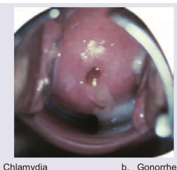
Identify the STD. (Recent Neet Pattern 2016-17)

All are true about the image shown EXCEPT:

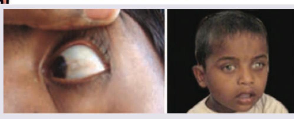
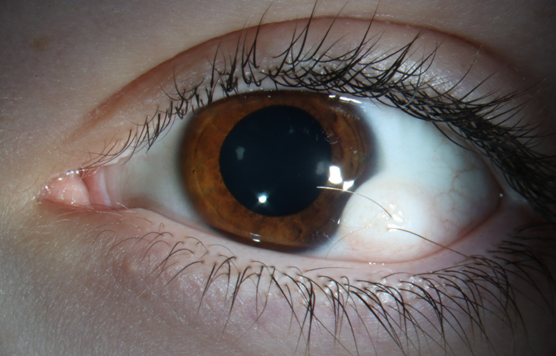
Identify the diagnosis based on the clinical image shown.

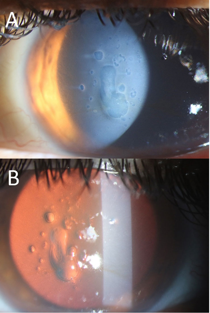
A patient, three years post-surgery, presents with the condition shown in the image and complains of decreased vision. What is the most likely diagnosis?

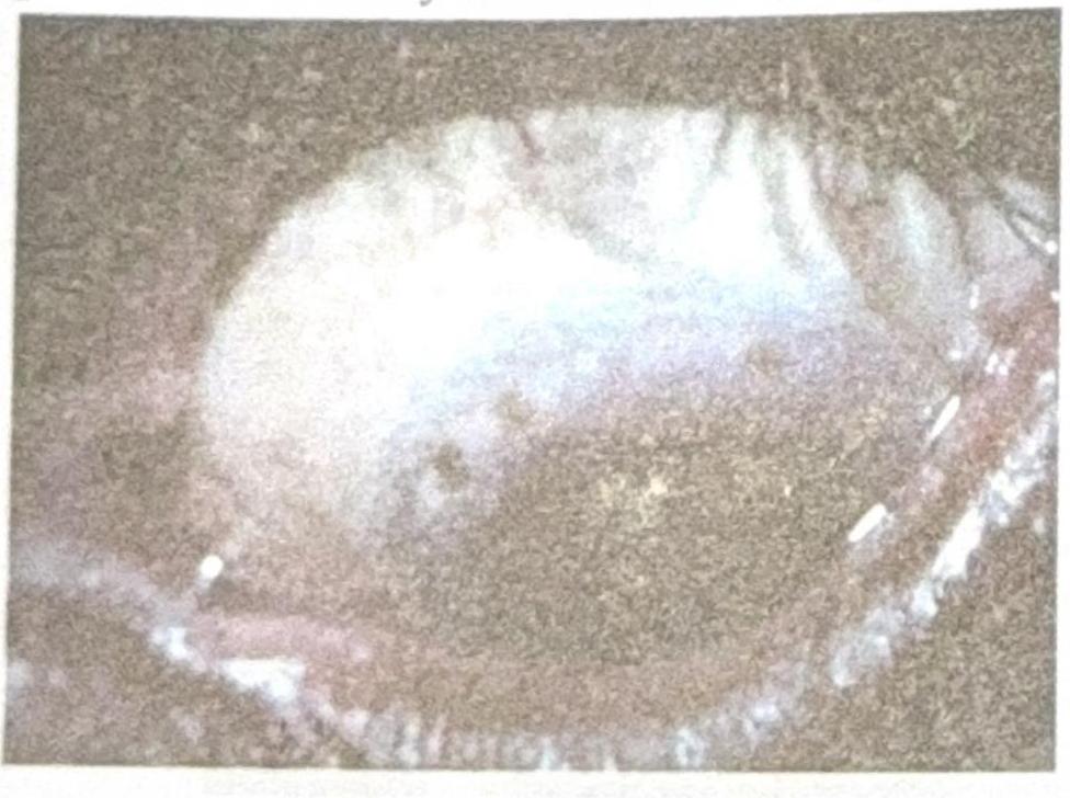
A patient presents to the OPD with the finding shown in the image. What is the most likely diagnosis?

Which of the following is not a feature of keratoconus?
Identify the correct sequence of staining in dry eyes? 1. Fluorescein stain 2. Lissamine green 3. Rose Bengal stain
Fleischer's ring is seen in?
True about Mooren's ulcer:
Cornea derives its nutrition chiefly from:
Explanation: ***Chlamydia*** - The image exhibits **cervicitis** with a visible **purulent exudate** and **cervical edema**, which is highly characteristic of **Chlamydia trachomatis** infection. - Given that Chlamydia is often **asymptomatic** or presents with non-specific symptoms, direct visualization of such changes on the cervix is a key diagnostic indicator in symptomatic cases. *Gonorrhea* - While gonorrhea can also cause cervicitis with purulent discharge, the specific appearance in the image with more pronounced **edema** and somewhat **friable cervix** is more classically associated with Chlamydia in many clinical presentations. - Differentiation often requires **laboratory testing** (e.g., NAAT) as visual signs can overlap, but the depicted features lean more towards Chlamydia. *Syphilis* - Syphilis is typically characterized by **chancres** (painless ulcers) in its primary stage, or rashes and condylomata lata in secondary syphilis, none of which are seen in this image. - The image shows inflammation and discharge, not ulcerative lesions. *Chancroid* - Chancroid is characterized by **painful genital ulcers** with a characteristic soft base and often associated with **buboes** (swollen lymph nodes). - The findings in the image—cervical inflammation and discharge—do not align with the typical presentation of chancroid.
Explanation: ***Plasma retinol level is the most accurate indicator of Vitamin A status*** - While plasma retinol levels can provide an indication of Vitamin A status, they are **not the most accurate** indicator, especially in marginal deficiency. The body tightly regulates plasma retinol levels until liver stores are severely depleted, meaning a normal plasma level does not rule out subclinical deficiency. - **Liver biopsy** for direct measurement of Vitamin A stores is considered the gold standard, though it is invasive. **Conjunctival Impression Cytology** (CIC) is another functional test useful for assessing corneal and conjunctival changes related to vitamin A deficiency. *Corynebacterium xerosis in cheesy plaques* - **Bitot's spots**, the cheesy plaques seen in the image, are accumulations of desquamated keratinized epithelial cells and mucus, often colonized by **Corynebacterium xerosis**. - This finding is a clinical sign of **Vitamin A deficiency**. *Squamous metaplasia* - In **Vitamin A deficiency**, the normal columnar epithelium of the conjunctiva undergoes **squamous metaplasia**, transforming into stratified squamous keratinizing epithelium. - This change contributes to the formation of Bitot's spots and can lead to **xerosis conjunctivae**. *Punctuate keratitis* - **Punctuate keratitis** can be a feature of **xerophthalmia**, the constellation of ocular signs due to Vitamin A deficiency. - It refers to dot-like epithelial lesions on the cornea, which can progress to more severe corneal involvement.
Explanation: ***Herbert's pits*** - The image displays characteristic **pits at the limbus**, which are a hallmark of healed follicular conjunctivitis, specifically **Herbert's pits**. - These pits are pathognomonic for **trachoma**, a chronic keratoconjunctivitis caused by *Chlamydia trachomatis*. *Horner-Trantas spots* - These are small, raised, gelatinous white dots found at the limbus, typically containing **eosinophils**. - They are characteristic of **vernal keratoconjunctivitis**, an allergic inflammatory condition, and not seen in the provided image. *Pannus* - **Pannus** refers to the growth of **vascularized connective tissue** onto the cornea. - While pannus is also a feature of trachoma, the image specifically shows distinct limbal pits, rather than diffuse vascularization. *Corneal dystrophy* - **Corneal dystrophies** are a group of inherited, bilateral, and progressive corneal disorders that manifest as opacities or structural changes in the cornea. - The findings in the image, such as pits, are inflammatory in origin and distinct from the typically stromal or epithelial changes seen in corneal dystrophies.
Explanation: ***Posterior Capsular Opacification*** - The image (especially part B, the **red reflex view**) shows a cloudy membrane with **Elschnig pearls** and fibrous tissue behind the intraocular lens, which is characteristic of **PCO**. - PCO commonly occurs months to years after cataract surgery and causes **decreased vision** due to scattering of light. *Corneal Dystrophy* - Corneal dystrophies primarily affect the **cornea**, leading to various forms of corneal opacification, and would typically appear as cloudiness or irregularities within the corneal layers. - While it can cause decreased vision, the opacification seen in the image is clearly behind the iris plane and involves the posterior capsule, not the cornea. *Pseudophakic Bullous Keratopathy* - This condition involves **corneal edema** and **bullae formation** due to endothelial cell dysfunction, leading to a hazy cornea. - The image does not show corneal edema or bullae; instead, the primary pathology is located at the **posterior capsule** of the intraocular lens. *Posterior Subcapsular Cataract (PSC)* - A PSC is a type of cataract that forms **before cataract surgery** and affects the natural crystalline lens. - The patient is three years post-surgery, indicating that the natural lens has been replaced, making a PSC (of the natural lens) an impossible diagnosis in this context.
Explanation: ***Limbal dermoid*** - A **limbal dermoid** is a congenital benign tumor typically found at the **limbus**, the junction between the cornea and sclera. - It appears as a **raised, yellowish-white mass** and may contain elements like hair follicles or sebaceous glands. *Pyogenic granuloma* - A **pyogenic granuloma** is a rapidly growing, benign vascular lesion that typically occurs after trauma or surgery. - It presents as a **red, pedunculated nodule** that bleeds easily, unlike the described finding. *Papilloma* - **Papillomas** are benign epithelial tumors caused by the human papillomavirus (HPV). - They appear as **small, fleshy growths** with a cauliflower-like surface and are less common at the limbus. *Pinguecula* - A **pinguecula** is a common, benign yellowish growth on the conjunctiva, usually located on the nasal side of the cornea. - It does not typically involve the **cornea** or appear as a raised mass like a dermoid tumor.
Explanation: ***Haab's striae*** - **Haab's striae** are characteristic of congenital glaucoma, presenting as horizontal breaks in Descemet's membrane due to increased intraocular pressure and corneal stretching. - In contrast, keratoconus involves **vertical breaks in Descemet's membrane** (due to corneal thinning and protrusion), known as Vautin's striae or Vogt's striae, not Haab's striae. *Irregular astigmatism* - **Irregular astigmatism** is a hallmark of keratoconus, resulting from the asymmetric and non-uniform thinning and protrusion of the cornea. - This irregular corneal shape leads to a significant reduction in visual acuity that cannot be fully corrected with spherical or regular cylindrical lenses. *Scissoring reflex* - The **scissoring reflex** (or scissoring phenomena) is an objective finding observed during retinoscopy in patients with keratoconus, indicating significant irregular astigmatism. - It refers to the opposing movements of the retinoscopic reflex, resembling the opening and closing of scissors, as the light across the pupil is neutralized. *Munson's sign* - **Munson's sign** is a classic clinical finding in advanced keratoconus, where the lower eyelid protrudes in a V-shape when the patient looks downwards. - This protrusion is caused by the conical shape of the cornea pushing against the eyelid.
Explanation: ***iii, ii, i*** - The correct order for staining in dry eyes is **Rose Bengal, Lissamine Green, and then Fluorescein**. This sequence minimizes the discomfort and potential interference between the dyes. - **Rose Bengal** and **Lissamine Green** stain devitalized or damaged epithelial cells and mucus, while **Fluorescein** stains areas where the epithelium is absent, indicating corneal or conjunctival abrasions or erosions. *i, iii, ii* - This sequence is incorrect because applying fluorescein first could mask the interpretation of epithelial damage by Lissamine Green or Rose Bengal, as it highlights epithelial defects rather than devitalized cells. - The dyes should be applied in an order that optimizes the visualization of different aspects of ocular surface health. *i, ii, iii* - Applying **Fluorescein** first is generally not recommended as it can spread over the ocular surface and potentially interfere with the specific staining patterns of other vital dyes like Lissamine Green or Rose Bengal. - This order does not follow the standard clinical practice for efficient and accurate dry eye assessment. *ii, iii, i* - While Lissamine Green can be used before Fluorescein, placing Rose Bengal after Lissamine Green but still before Fluorescein is not the optimal sequence. - The standard practice aims to assess different layers or types of damage sequentially for a comprehensive evaluation.
Explanation: ***Keratoconus*** - **Fleischer's ring** is a brownish-yellow to greenish-brown ring of **iron deposition** (hemosiderin) in the **corneal epithelium** around the base of the cone in patients with **keratoconus**. - It is a specific sign of **keratoconus**, which is a progressive thinning and steepening of the cornea, causing it to bulge into a cone shape. *Keratoglobus* - **Keratoglobus** is a rare, non-inflammatory condition where the **entire cornea** becomes thin and globularly protrudes, unlike the localized cone in keratoconus. - It typically does not present with an isolated Fleischer's ring; other corneal findings may be present. *Keratomalacia* - **Keratomalacia** is corneal softening and necrosis caused by severe **vitamin A deficiency**. - It presents with xerosis (dryness), night blindness, and eventual corneal melting and perforation. - It does not involve iron deposition in the form of a Fleischer's ring. *Trachoma* - **Trachoma** is a chronic infectious disease of the eye caused by *Chlamydia trachomatis*, leading to scarring of the conjunctiva and cornea. - It is a leading cause of preventable blindness worldwide and is not associated with Fleischer's ring.
Explanation: ***Affects cornea*** - **Mooren's ulcer** is a rare, severe, and idiopathic chronic **ulcerative keratitis** that primarily affects the cornea. - It involves a progressive **peripheral corneal stromal thinning** and ulceration, often extending circumferentially and centrally. *Bilateral in majority of cases* - Mooren's ulcer is **unilateral in approximately 60-80%** of cases, making bilateral presentation less common. - While it can be bilateral, especially in a more severe form, it's not the majority presentation. *Sudden loss of vision* - The vision loss associated with Mooren's ulcer is typically **gradual and progressive**, due to increasing corneal thinning, scarring, and astigmatism. - **Sudden vision loss** is not a characteristic feature of this condition and would suggest other acute pathologies. *Painless* - Mooren's ulcer is often associated with **significant pain**, **photophobia**, and **lacrimation**. - The pain can be severe and debilitating, stemming from the inflammatory process and corneal nerve involvement.
Explanation: ***Aqueous humour*** - The cornea is **avascular**, meaning it lacks its own blood vessels, and thus relies on the **aqueous humor** for metabolic support. - The aqueous humor, located in the anterior chamber, provides **glucose, amino acids, and oxygen** to the posterior surface of the cornea. *Corneal vessels* - The **cornea is avascular** under normal physiological conditions; the presence of corneal vessels (neovascularization) is typically a sign of pathology. - This avascularity is crucial for maintaining corneal transparency and optimal vision. *Perilimbal vessels* - Perilimbal vessels (at the limbus, the junction between cornea and sclera) primarily supply the **periphery of the cornea** and the limbal stem cell niche, not the entire cornea's main nutritional needs. - While they contribute some oxygen and nutrients, the **aqueous humor** is the predominant source for the central and posterior cornea. *Mucous layer* - The mucous layer, part of the tear film, primarily functions in **lubrication and adhesion** of the tear film to the corneal surface. - While it contains some dissolved oxygen from the atmosphere to supply the anterior cornea, its role in nutrient supply (glucose, amino acids) is **minimal** compared to the aqueous humor.
Corneal Anatomy and Physiology
Practice Questions
Bacterial Keratitis
Practice Questions
Viral Keratitis
Practice Questions
Fungal Keratitis
Practice Questions
Protozoan Keratitis
Practice Questions
Corneal Degenerations
Practice Questions
Corneal Dystrophies
Practice Questions
Keratoconus and Ectatic Disorders
Practice Questions
Corneal Transplantation
Practice Questions
Corneal Topography and Imaging
Practice Questions
Dry Eye Disease
Practice Questions
Corneal Trauma
Practice Questions
Get full access to all questions, explanations, and performance tracking.
Start For Free