
Diseases of the Cornea — MCQs

On this page
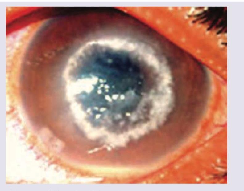
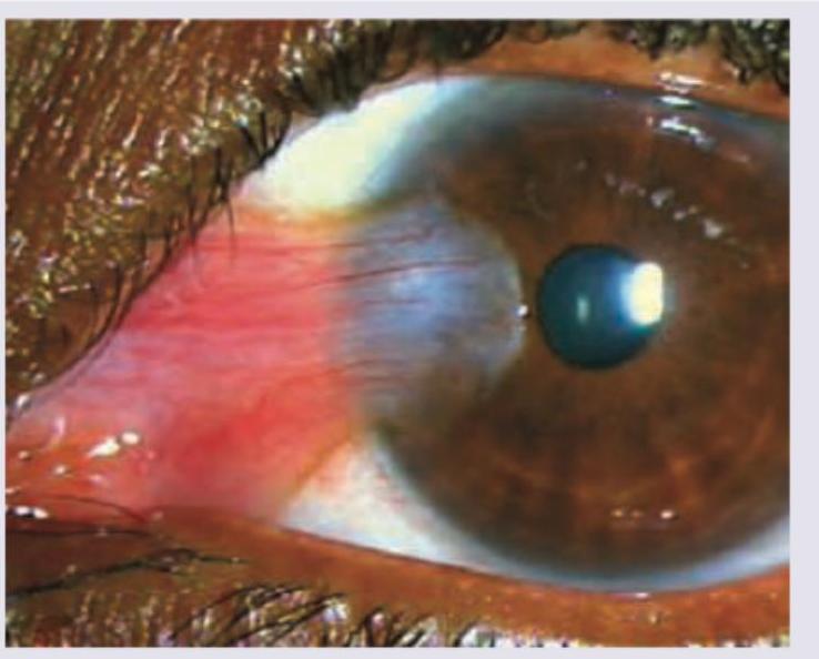
A patient presents with a painful red eye. Fluorescein staining reveals the lesion shown in the image. What is the diagnosis?
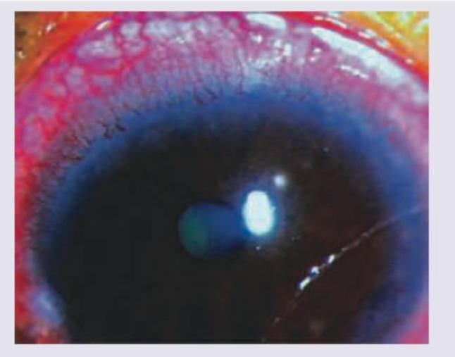
A 15-year-old contact lens user presents with severe eye pain for last 3 days. All are correct about the treatment for the condition shown in the patient except:
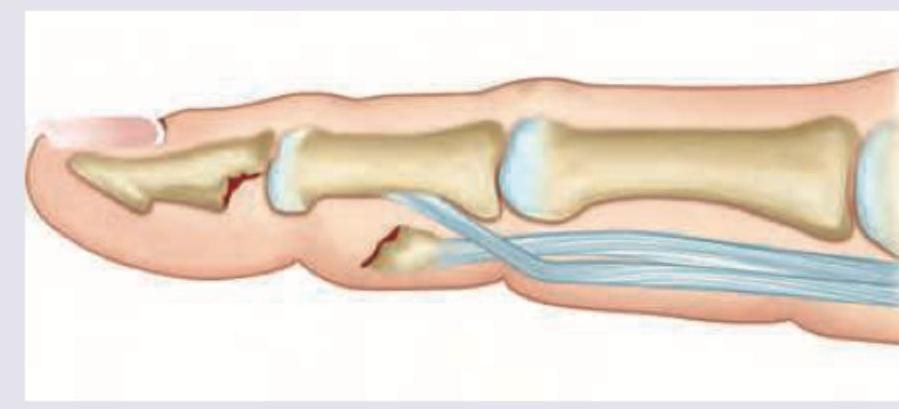
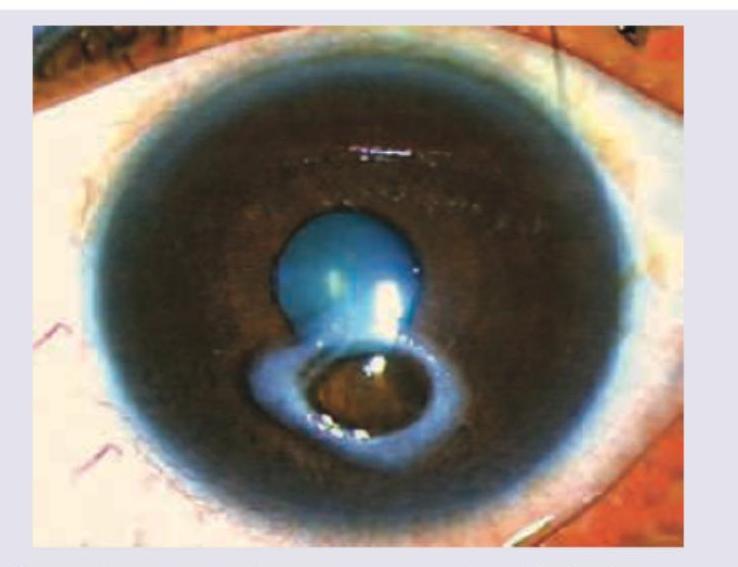
Which complication of the corneal ulcer is shown in the image below?
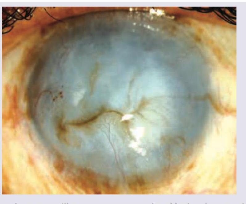
What does the following image show?
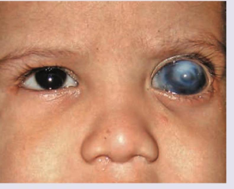
An 18-year-old male presents with complaints of acute pain and redness in the right eye with reduced visual acuity and a white spot on the cornea. What does the given ocular examination reveal?
All are correct about the lesion shown except:
What does the following image show?
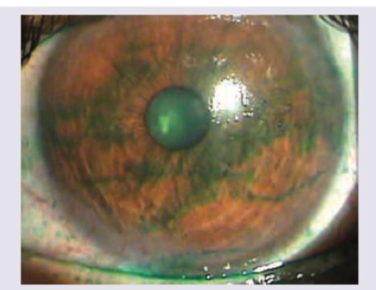
Identify the stain instilled in the eye in the following image:
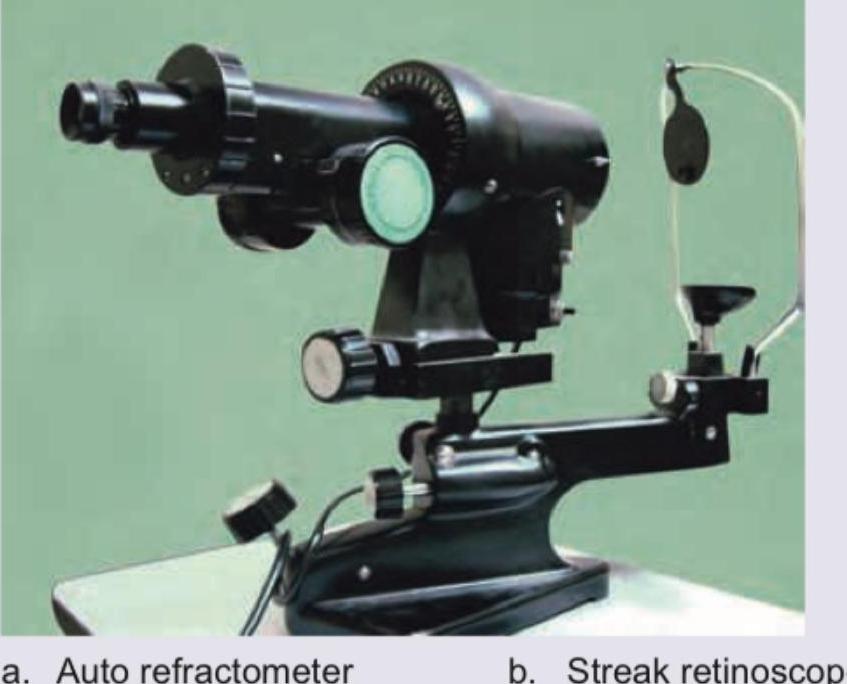
Name the instrument shown in the image:
All the following statements regarding the picture are true except: (Recent Neet Pattern 2016-17)
Practice by Chapter
Corneal Anatomy and Physiology
Practice Questions
Bacterial Keratitis
Practice Questions
Viral Keratitis
Practice Questions
Fungal Keratitis
Practice Questions
Protozoan Keratitis
Practice Questions
Corneal Degenerations
Practice Questions
Corneal Dystrophies
Practice Questions
Keratoconus and Ectatic Disorders
Practice Questions
Corneal Transplantation
Practice Questions
Corneal Topography and Imaging
Practice Questions
Dry Eye Disease
Practice Questions
Corneal Trauma
Practice Questions
Want unlimited practice?
Get full access to all questions, explanations, and performance tracking.
Scan to download app