What is the most common cause of keratitis in soft contact lens users?
Angioid streaks in the eyes are seen in which of the following conditions?
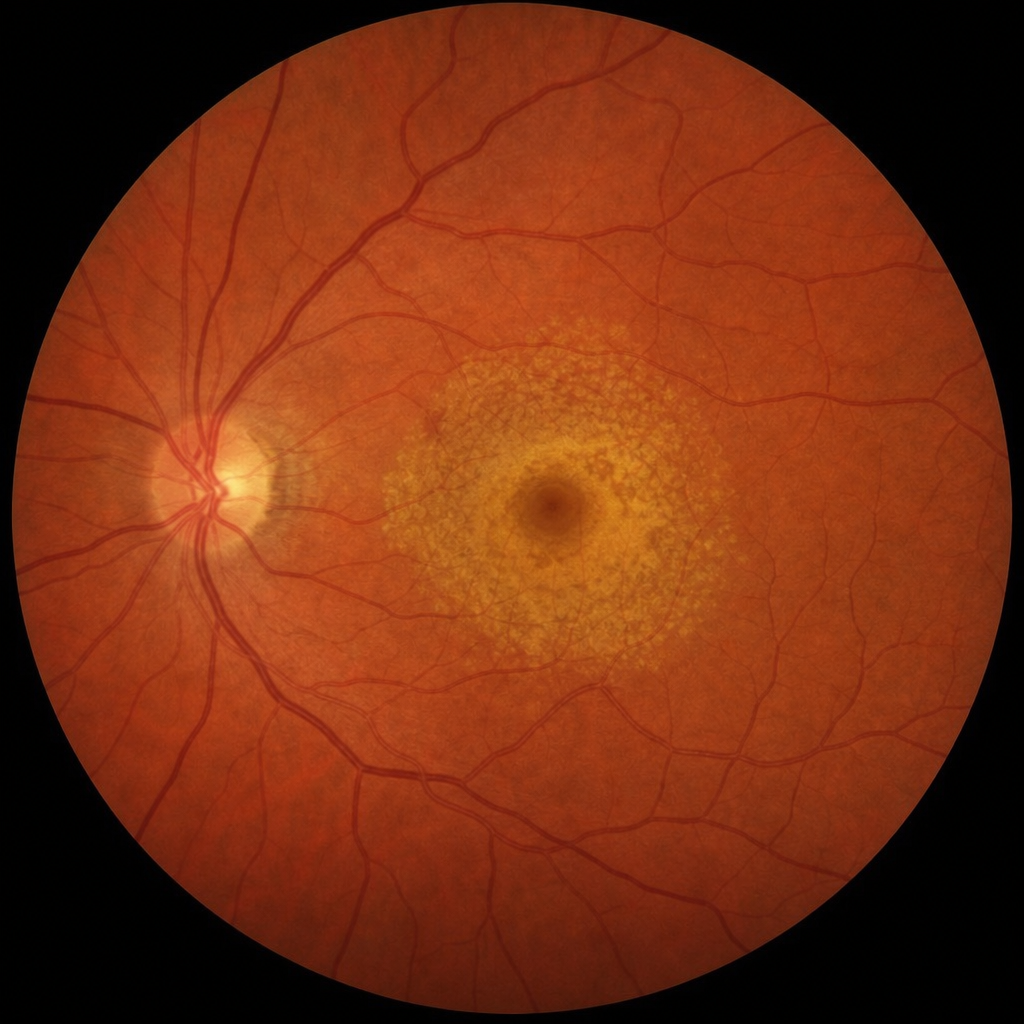
An otherwise healthy person who wears contact lenses develops a small ulceration of the eye. What is the most likely cause?
All are features of fungal keratitis, EXCEPT?
Which of the following statements is true regarding the endothelium of the cornea?
What is true about keratoconus?
Which of the following bacteria can invade intact corneal epithelium?
Radial keratoneuritis is a feature of which of the following conditions?
Which of the following drugs is used as an adjuvant therapy for the treatment of fungal corneal ulcers?
Which of the following methods is used to diagnose a corneal ulcer caused by Acanthamoeba?
Explanation: **Explanation:** **Acanthamoeba** is the most common cause of keratitis specifically associated with **soft contact lens** use, particularly when combined with poor hygiene practices such as using tap water for cleaning lenses, swimming while wearing lenses, or using contaminated lens cases. Acanthamoeba is a free-living soil and water amoeba that exists in two forms: the active trophozoite and the resilient double-walled cyst. The cysts are highly resistant to standard disinfection, making this infection difficult to treat. **Analysis of Options:** * **Acanthamoeba (Correct):** It is the classic association for contact lens-related keratitis. The hallmark clinical sign is a **ring-shaped corneal infiltrate** and pain out of proportion to clinical findings. * **Staphylococcus aureus:** While a common cause of bacterial keratitis generally, it is not the *most* specific or common association for contact lens users compared to *Pseudomonas aeruginosa* (bacterial) or *Acanthamoeba* (parasitic). * **Naegleria:** Although a free-living amoeba, it primarily causes Primary Amoebic Meningoencephalitis (PAM) and is not a standard cause of keratitis. * **Herpes simplex virus:** This is the most common cause of infectious corneal blindness in developed countries, typically presenting with **dendritic ulcers**, but its etiology is viral reactivation rather than contact lens wear. **High-Yield Clinical Pearls for NEET-PG:** * **Most common bacterial cause in CL users:** *Pseudomonas aeruginosa* (presents with rapid progression and greenish-yellow discharge). * **Pathognomonic sign of Acanthamoeba:** Radial keratoneuritis (infiltration along corneal nerves). * **Diagnosis:** Confirmed by culture on **Non-nutrient agar with E. coli overlay** or confocal microscopy. * **Treatment:** Topical biguanides (PHMB) or diamidines (Propamidine/Brolene).
Explanation: **Explanation:** **Angioid streaks** are irregular, jagged, radiating lines deep to the retina that resemble blood vessels. They represent **crack-like dehiscences (breaks) in a thickened, calcified Bruch’s membrane**, located between the retinal pigment epithelium and the choriocapillaris. 1. **Why Pseudoxanthoma Elasticum (PXE) is correct:** PXE (Grönblad-Strandberg syndrome) is the most common systemic association of angioid streaks. It is a genetic disorder characterized by the progressive calcification of elastic fibers in the skin, eyes, and cardiovascular system. In the eye, this calcification makes Bruch’s membrane brittle, leading to the characteristic streaks. 2. **Why the other options are incorrect:** * **Tendinous xanthoma, Xanthelasma, and Eruptive xanthoma** are all cutaneous manifestations of lipid metabolism disorders (hyperlipidemias). While they involve the deposition of cholesterol-rich material in tendons or skin (especially the eyelids in xanthelasma), they do not involve the calcification of Bruch’s membrane and are not associated with angioid streaks. **High-Yield Clinical Pearls for NEET-PG:** * **Mnemonic "PEPSI" for Angioid Streaks:** * **P** – **P**seudoxanthoma elasticum (Most common) * **E** – **E**hlers-Danlos syndrome * **P** – **P**aget’s disease of bone * **S** – **S**ickle cell anemia (and other hemoglobinopathies like Thalassemia) * **I** – **I**diopathic * **Clinical Significance:** The primary vision-threatening complication of angioid streaks is **Choroidal Neovascularization (CNV)**, which can lead to subretinal hemorrhage and macular scarring. * **Skin Finding in PXE:** Look for "plucked chicken skin" appearance on the neck or axilla.
Explanation: **Explanation:** The clinical scenario describes a contact lens wearer with a corneal ulcer, which is a classic presentation for **Acanthamoeba keratitis**. **1. Why Acanthamoeba is correct:** Acanthamoeba is a free-living amoeba found in soil and water. In the context of NEET-PG, the strongest risk factor for this infection is **contact lens use** (especially involving poor hygiene, such as using tap water for cleaning or swimming with lenses). The organism exists in two forms: the active trophozoite and the resilient cyst. A hallmark clinical feature is **pain out of proportion to clinical findings** and the presence of a **ring-shaped stromal infiltrate** in later stages. **2. Why the other options are incorrect:** * **Cytomegalovirus (CMV):** Typically causes retinitis (pizza-pie appearance) in immunocompromised patients (e.g., HIV/AIDS). It does not typically cause primary corneal ulcers in healthy individuals. * **Toxocara:** Usually presents as **Ocular Larva Migrans**, characterized by posterior pole granulomas or endophthalmitis, often in children who have contact with puppies. * **Toxoplasma:** The most common cause of posterior uveitis (retinochoroiditis). It presents with a "headlight in the fog" appearance on fundoscopy, not a corneal ulcer. **High-Yield Clinical Pearls for NEET-PG:** * **Stain of choice:** Calcofluor white (binds to cysts) or Periodic acid-Schiff (PAS). * **Culture medium:** Non-nutrient agar enriched with *E. coli*. * **Early sign:** Radial keratoneuritis (infiltration along corneal nerves). * **Treatment:** Topical biguanides (PHMB) or Chlorhexidine.
Explanation: **Explanation:** Fungal keratitis (Keratomycosis) is a common cause of corneal ulceration, particularly in agricultural workers following trauma with vegetative matter. **Why "Early vascularisation" is the correct answer:** Fungal keratitis is characterized by a **lack of early vascularization**. Fungi produce certain toxins and enzymes that suppress the inflammatory response and inhibit the ingrowth of new blood vessels. In contrast, bacterial ulcers tend to induce neovascularization much earlier. If vascularization occurs in fungal cases, it is usually a very late feature during the healing phase. **Analysis of other options:** * **A. Less symptoms, more signs:** This is a hallmark of fungal keratitis. Due to the slow-growing nature of fungi and the relative lack of acute inflammatory toxins compared to bacteria, the patient’s pain and discomfort are often disproportionately mild compared to the extensive clinical findings (large ulcer, hypopyon). * **B. Infected hypopyon:** Fungi can penetrate an intact Descemet’s membrane without perforation. This leads to an "infected" hypopyon (containing actual fungal elements), unlike the "sterile" hypopyon seen in bacterial ulcers. * **D. Dry white slough:** Fungal ulcers typically present as a dry, grayish-white, elevated lesion with "feathery" finger-like extensions into the surrounding stroma. **Clinical Pearls for NEET-PG:** * **Most common cause:** *Aspergillus* (overall), *Fusarium* (after trauma), *Candida* (in pre-existing ocular surface disease). * **Key Signs:** Satellite lesions (small infiltrates around the main ulcer), Immune ring (Wessely ring), and a fixed, non-mobile hypopyon. * **Diagnosis:** Potassium Hydroxide (KOH) mount is the initial investigation; Sabouraud’s Dextrose Agar (SDA) is the culture medium of choice. * **Treatment:** Topical Natamycin (5%) is the drug of choice for filamentous fungi.
Explanation: **Explanation:** The corneal endothelium is a single layer of hexagonal cells that plays a vital role in maintaining corneal transparency. **Why Option C is Correct:** The cornea is naturally inclined to absorb water from the aqueous humor due to the swelling pressure of the stroma. The endothelium maintains the cornea in a state of **relative dehydration (deturgescence)** through two mechanisms: 1. **Mechanical Barrier:** It acts as a semi-permeable barrier. 2. **Active Na+/K+ ATPase Pump:** This is the "Endothelial Pump" that actively transports ions out of the stroma into the aqueous humor, with water following osmotically. This prevents corneal edema and maintains clarity. **Why Other Options are Incorrect:** * **Option A:** The endothelium is a **monolayer** (single layer) of cells, not three layers. * **Option B:** Human corneal endothelial cells have **no regenerative capacity**. Once damaged, they do not undergo mitosis. Instead, neighboring cells enlarge (pleomorphism) and spread out (polymegathism) to cover the defect. * **Option D:** Endothelial cell density **decreases** with age and following intraocular surgeries (like cataract surgery). The normal adult count is approximately 2500–3000 cells/mm². **High-Yield Clinical Pearls for NEET-PG:** * **Critical Threshold:** If the endothelial cell count falls below **500 cells/mm²**, the pump fails, leading to irreversible corneal edema (Bullous Keratopathy). * **Specular Microscopy:** The gold standard investigation to study endothelial cell morphology and count. * **Pachymetry:** Used to measure corneal thickness; an increase in thickness is often the first sign of endothelial dysfunction. * **Fuchs' Endothelial Dystrophy:** A common condition characterized by "guttae" (droplet-like excrescences) and progressive loss of endothelial cells.
Explanation: ### Explanation **Keratoconus** is a non-inflammatory, progressive ectatic disorder characterized by **progressive thinning of the corneal stroma**, usually starting at puberty. This thinning causes the cornea to bulge into a conical shape due to intraocular pressure, leading to irregular astigmatism and high myopia. **Analysis of Options:** * **A (Correct):** The hallmark of keratoconus is the progressive thinning and protrusion of the central or paracentral cornea. * **B (Incorrect):** While **Fleischer rings** (iron deposits at the base of the cone) are a classic sign of keratoconus, the question asks for the most fundamental definition. In many NEET-PG style questions, if a pathological process (thinning) is pitted against a clinical sign (ring), the underlying pathology is the "more true" or defining characteristic. *Note: In some exams, this might be considered a multiple-correct scenario, but "progressive thinning" is the definitive physiological change.* * **C (Incorrect):** Soft contact lenses **are used** in very early/mild stages. As the disease progresses, RGP (Rigid Gas Permeable) or Scleral lenses become necessary to provide a regular refracting surface. * **D (Incorrect):** Keratoconus typically causes **progressive myopia** and irregular astigmatism, not hyperopia. **High-Yield Clinical Pearls for NEET-PG:** * **Early Signs:** Frequent changes in spectacle power and "scissors reflex" on retinoscopy. * **Advanced Signs:** * **Munson’s Sign:** V-shaped indentation of the lower lid on downgaze. * **Vogt’s Striae:** Vertical stress lines in the deep stroma. * **Rizutti’s Sign:** Conical reflection on the nasal cornea when light is shone from the temporal side. * **Acute Hydrops:** Sudden corneal edema due to a rupture in **Descemet’s membrane**. * **Treatment:** **CXL (Corneal Collagen Cross-linking)** with Riboflavin and UVA is the only treatment to stop progression. Penetrating keratoplasty is the gold standard for advanced cases with scarring.
Explanation: ### Explanation The corneal epithelium acts as a formidable mechanical barrier against most pathogens. Most bacteria require a pre-existing epithelial defect (trauma, contact lens wear, or dry eye) to initiate an infection. However, a specific group of organisms possesses virulence factors that allow them to penetrate and invade an **intact** corneal epithelium. **Correct Answer: D. Legionella** While *Legionella* is primarily known for causing pneumonia (Legionnaires' disease), it is classically listed in ophthalmic literature as one of the rare organisms capable of penetrating intact corneal epithelium. This is a high-yield fact often tested in postgraduate entrance exams to distinguish it from common ocular pathogens. **Analysis of Incorrect Options:** * **A, B, and C (Neisseria gonorrhoeae, Corynebacterium diphtheriae, and Neisseria meningitidis):** These are actually the **other** primary members of the group that can invade intact epithelium. In the context of this specific question format (where D is marked correct), it implies a "Which of the following" selection where Legionella is the intended answer, though typically, options A, B, and C are the most frequently cited examples in standard textbooks like Khurana. **High-Yield Clinical Pearls for NEET-PG:** To remember the organisms that can penetrate an intact cornea, use the mnemonic **"CHLNL"** or **"NHL"**: 1. ***C*orynebacterium diphtheriae** 2. ***H*aemophilus aegyptius** (Koch-Weeks bacillus) 3. ***L*isteria monocytogenes** 4. ***N*eisseria gonorrhoeae** and ***N*eisseria meningitidis** 5. ***L*egionella** species 6. ***S*higella** (rarely cited) * **Most common cause of bacterial keratitis overall:** *Staphylococcus aureus*. * **Most common cause in contact lens users:** *Pseudomonas aeruginosa*. * **Key Clinical Sign:** Invasion of intact epithelium often leads to rapid corneal perforation if not treated aggressively.
Explanation: **Explanation:** **Radial keratoneuritis** is a pathognomonic clinical feature of **Acanthamoeba keratitis**. It refers to the infiltration of corneal nerves by the *Acanthamoeba* trophozoites, appearing as linear, radial infiltrates extending from the limbus toward the center of the cornea. This perineural involvement explains the characteristic **disproportionate pain** (pain much more severe than the clinical signs) experienced by these patients. * **Acanthamoeba keratitis (Correct):** Typically occurs in contact lens users (80% of cases) who use contaminated tap water or swim while wearing lenses. Early signs include "pseudodendrites," followed by radial keratoneuritis, and eventually a classic **ring-shaped stromal infiltrate**. * **Herpes zoster keratitis (Incorrect):** Characterized by pseudodendrites (stuck-on appearance without terminal bulbs) and decreased corneal sensations, but does not feature radial keratoneuritis. * **Neuroparalytic keratitis (Incorrect):** Occurs due to damage to the Trigeminal nerve (CN V), leading to loss of sensation and subsequent epithelial breakdown; it is a degenerative rather than an infiltrative process. **High-Yield Clinical Pearls for NEET-PG:** * **Gold Standard Diagnosis:** Culture on **Non-nutrient agar with E. coli overlay**. * **Stains:** Calcofluor white (fluorescent) or Periodic acid-Schiff (PAS). * **Treatment of Choice:** Topical biguanides (e.g., **PHMB** 0.02% or **Chlorhexidine**) and diamidines (e.g., Propamidine/Brolene). * **Differential Diagnosis:** Often misdiagnosed as Fungal or Herpetic keratitis due to its slow progression and resistance to standard antibiotics.
Explanation: **Explanation:** In the management of infectious keratitis (including fungal corneal ulcers), **Atropine (1%) eye drops** serve as a vital adjuvant therapy. The primary medical rationale for its use is to manage **secondary anterior uveitis** and **ciliary muscle spasm**, which are common complications of deep corneal ulcers. **Why Atropine is the Correct Answer:** 1. **Cycloplegia:** It paralyzes the ciliary muscle, relieving the intense "ciliary spasm" pain. 2. **Mydriasis:** It dilates the pupil, preventing the formation of **posterior synechiae** (adhesion of the iris to the lens). 3. **Reduced Inflammation:** By causing vasoconstriction of the ciliary body vessels, it reduces the exudation of inflammatory cells into the anterior chamber. **Analysis of Incorrect Options:** * **Pilocarpine (Option B):** This is a miotic. It increases ciliary muscle contraction and inflammation, which would worsen the pain and promote the formation of small, fixed pupils (annular synechiae). * **Dexamethasone (Option C):** Corticosteroids are generally **contraindicated** in active fungal ulcers. They inhibit local immunity and promote fungal replication and "melting" of the cornea (keratomalacia), potentially leading to perforation. * **Lidocaine (Option D):** While a local anesthetic, it is not used as a treatment adjuvant. Repeated use of topical anesthetics is toxic to the corneal epithelium and can delay healing. **High-Yield Clinical Pearls for NEET-PG:** * **Drug of Choice (DOC) for Fungal Keratitis:** Topical **Natamycin (5%)** is the first-line treatment for filamentous fungi (e.g., *Aspergillus, Fusarium*). * **Characteristic Sign:** Fungal ulcers often present with **"Satellite lesions"** and a dry, feathery edge. * **Atropine Duration:** It is the strongest cycloplegic, with effects lasting 7–10 days. In children, watch for systemic toxicity (dry mouth, flushing, tachycardia).
Explanation: **Explanation:** Acanthamoeba keratitis is a sight-threatening infection often associated with contact lens wear or exposure to contaminated water. Diagnosing it requires identifying the characteristic double-walled cysts or trophozoites in corneal scrapings. **Why "All of the above" is correct:** While Acanthamoeba is a protozoan, its cell wall contains chitin and cellulose, making it susceptible to stains and methods typically used for fungi: * **KOH Mount (10%):** This is a rapid bedside technique. KOH dissolves cellular debris, allowing the highly resistant, double-walled Acanthamoeba cysts to be visualized under a light microscope. * **Calcofluor White:** This is a fluorescent stain that binds to cellulose and chitin. It is considered highly sensitive for identifying Acanthamoeba cysts, which appear as bright apple-green or blue-white structures under a fluorescence microscope. * **Lactophenol Cotton Blue (LPCB):** Though primarily a fungal stain, LPCB can be used to stain the cysts, providing contrast to visualize their morphology. **High-Yield Clinical Pearls for NEET-PG:** * **Gold Standard Culture:** Non-nutrient agar (NNA) enriched with **Escherichia coli** (look for "trailing" or "tracks" made by amoeba). * **Classic Clinical Sign:** **Ring Infiltrate** (advanced stage) and **Radial Keratoneuritis** (early stage, pathognomonic). * **Symptoms vs. Signs:** Characterized by **pain out of proportion** to the clinical findings. * **Other Stains:** Periodic Acid-Schiff (PAS) and Giemsa stain can also be used. * **Treatment of Choice:** Topical biguanides (PHMB 0.02%) or Diamidines (Propamidine isethionate/Brolene).
Corneal Anatomy and Physiology
Practice Questions
Bacterial Keratitis
Practice Questions
Viral Keratitis
Practice Questions
Fungal Keratitis
Practice Questions
Protozoan Keratitis
Practice Questions
Corneal Degenerations
Practice Questions
Corneal Dystrophies
Practice Questions
Keratoconus and Ectatic Disorders
Practice Questions
Corneal Transplantation
Practice Questions
Corneal Topography and Imaging
Practice Questions
Dry Eye Disease
Practice Questions
Corneal Trauma
Practice Questions
Get full access to all questions, explanations, and performance tracking.
Start For Free