
Diseases of the Cornea — MCQs

On this page
Hypopyon corneal ulcer is caused by which microorganism?
Which of the following is the least common corneal dystrophy?
Which of the following is not an indication for keratoplasty?
Not true regarding herpes keratitis?
A 48-year-old diabetic with orbital cellulitis presented with a corneal ulcer. An aqueous tap showed branched hyphae. What is the diagnosis?
Cloudy cornea is seen in which of the following conditions?
Deposition of which of the following is seen in the Descemet's membrane in Kayser-Fleischer rings?
All of the following are true regarding keratoconus except?
What is the toughest layer of the cornea?
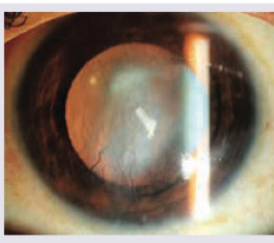
In the given condition, what is the cause of defective vision?
Practice by Chapter
Corneal Anatomy and Physiology
Practice Questions
Bacterial Keratitis
Practice Questions
Viral Keratitis
Practice Questions
Fungal Keratitis
Practice Questions
Protozoan Keratitis
Practice Questions
Corneal Degenerations
Practice Questions
Corneal Dystrophies
Practice Questions
Keratoconus and Ectatic Disorders
Practice Questions
Corneal Transplantation
Practice Questions
Corneal Topography and Imaging
Practice Questions
Dry Eye Disease
Practice Questions
Corneal Trauma
Practice Questions
Want unlimited practice?
Get full access to all questions, explanations, and performance tracking.
Scan to download app