All are true regarding cornea except:
Sensory innervation of the cornea is by which nerve?
Density of cells in adult corneal endothelium is
Conjunctival epithelium is a type of what?
Which among the following is the BEST irrigating fluid during ECCE?
A 20-year-old male complains of repeated changes in glasses prescription. This is most likely caused by:
In uveitis, site of keratic precipitate is:
Which of the following statements about Fuchs' corneal dystrophy is true?
What does a visual acuity test primarily assess?
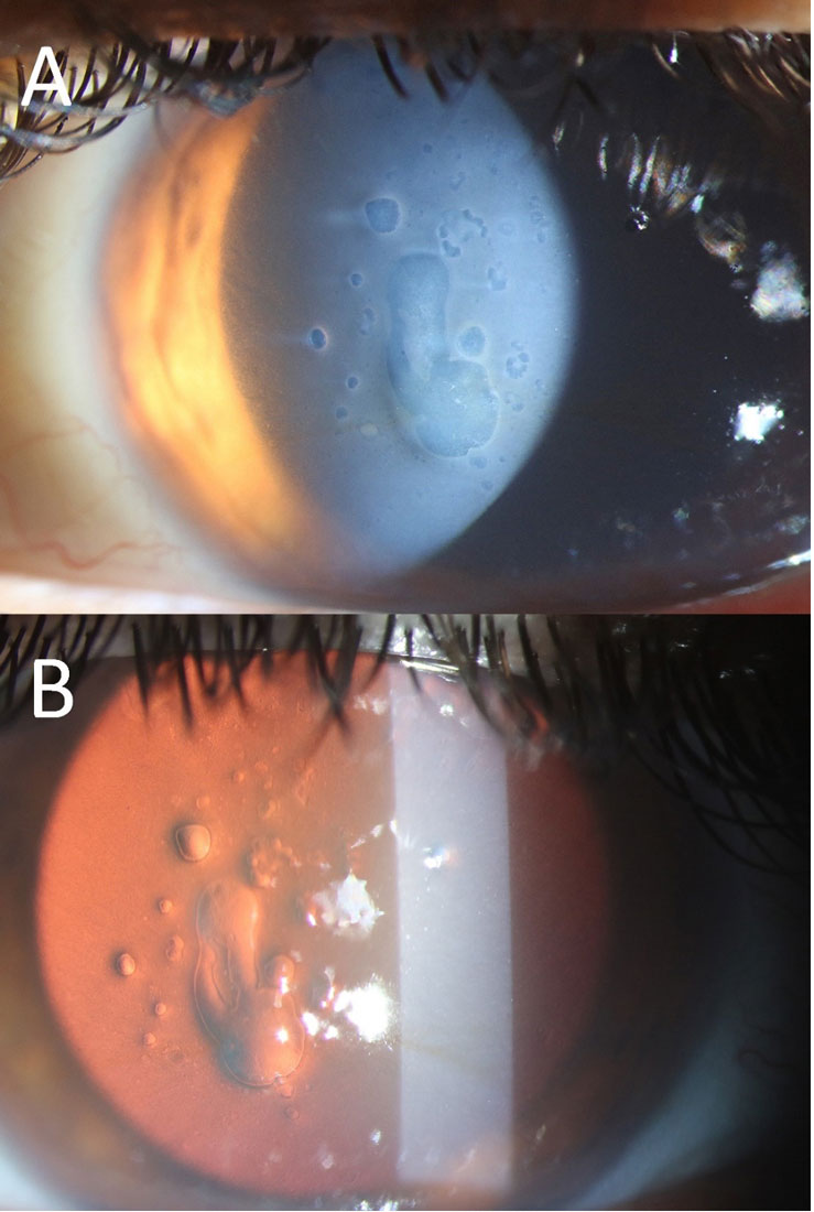
A patient, three years post-surgery, presents with the condition shown in the image and complains of decreased vision. What is the most likely diagnosis?

Explanation: ***The cornea is richly vascular.*** - The cornea is an **avascular** tissue, meaning it lacks blood vessels, which is crucial for its transparency. - This avascularity helps prevent light scatter and maintains clear vision. - The cornea receives nutrition from the **aqueous humor**, **tear film**, and **limbal blood vessels** through diffusion. *Endothelium help in maintaining dehydrated state* - The **corneal endothelium** actively pumps fluid out of the corneal stroma via **Na-K-ATPase pumps**, preventing swelling and maintaining its relative state of dehydration. - This **deturgescence** is essential for the cornea's transparency and optimal refractive power. *Oxygen is mostly derived by epithelium directly from the air through tear film* - The corneal **epithelium** primarily obtains oxygen directly from the atmosphere via the **tear film** when the eyes are open. - During sleep, oxygen is primarily supplied by the **palpebral conjunctival vessels**. *Corneal thickness is greater at periphery than center* - The cornea is **thinner at the center** (around 520-540 µm) and gradually **thicker towards the periphery** (around 600-640 µm). - This structural difference contributes to its optical properties and mechanical stability.
Explanation: ***5th (ophthalmic division of the trigeminal nerve)*** - The **trigeminal nerve (CN V)** is responsible for sensory innervation of the face, and its **ophthalmic division (V1)** specifically supplies the cornea. - This extensive sensory innervation explains the **extreme sensitivity of the cornea** to touch, temperature, and chemicals, and is crucial for the **corneal reflex**. *6th (Abducens nerve)* - The **abducens nerve (CN VI)** is a **motor nerve** responsible for innervating the **lateral rectus muscle**, which abducts the eye. - It has no role in the sensory innervation of the cornea. *3rd (Oculomotor nerve)* - The **oculomotor nerve (CN III)** is primarily a **motor nerve** responsible for innervating most of the **extraocular muscles** (superior, inferior, medial recti, inferior oblique) and the **levator palpebrae superioris**. - It also carries **parasympathetic fibers** for pupillary constriction and accommodation, but it does not provide sensory innervation to the cornea. *4th (Trochlear nerve)* - The **trochlear nerve (CN IV)** is a **motor nerve** that innervates the **superior oblique muscle**, which depresses and internally rotates the eye. - It has no function in corneal sensation.
Explanation: ***3000 cells/mm2*** - The **normal density** of corneal endothelial cells in a young adult is approximately **3000-3500 cells/mm²**. - This density is crucial for maintaining corneal clarity through its **pump function**. *2000 cells/mm2* - A density of **2000 cells/mm²** or lower in the corneal endothelium indicates a significantly reduced cell count. - This level is often considered the **minimum threshold** below which the cornea may lose its ability to remain clear, leading to **corneal edema**. *4000 cells/mm2* - While some individuals, especially younger ones, might have slightly higher densities, **4000 cells/mm²** is generally above the typical average for an adult. - This higher density is more common in **infants and young children**, where cell count is higher and gradually declines with age. *5000 cells/mm2* - A cell density of **5000 cells/mm²** is significantly higher than the normal adult range and is usually observed only in **neonates** or very young infants. - Such high densities are indicative of a developing or very young endothelium, not a typical adult state.
Explanation: ***Stratified non keratinised squamous*** - The **conjunctival epithelium** is primarily **stratified squamous epithelium**, meaning it has multiple layers of flattened cells. - It is **non-keratinized**, indicating that it does not produce keratin, which helps keep the surface moist and flexible. *Pseudostratified* - **Pseudostratified epithelium** appears to have multiple layers due to the varying heights of cells and nuclei at different levels, but all cells are in contact with the basement membrane. - A common example is in the **trachea**, where it is usually ciliated with goblet cells, not typically found in the conjunctiva. *Stratified columnar* - **Stratified columnar epithelium** has multiple layers of cells, with the outermost layer consisting of columnar cells. - This type of epithelium is relatively rare, found in certain ducts and parts of the urethra, and is not characteristic of the conjunctiva. *Transitional* - **Transitional epithelium**, also known as urothelium, is a specialized stratified epithelium found in the **urinary tract** (e.g., bladder, ureters). - It is unique for its ability to **stretch** and change shape, which is not a primary function of the conjunctival surface.
Explanation: ***Balanced salt solution + glutathione*** - **Balanced salt solution with glutathione** is considered the best irrigating fluid for ECCE because it closely mimics the **natural aqueous humor**, maintaining corneal endothelial cell health and viability during surgery. - The addition of **glutathione** provides an antioxidant effect, protecting the corneal endothelium from oxidative stress and maintaining its metabolic function during prolonged irrigation. *Ringer lactate* - While **Ringer's lactate** is a balanced electrolyte solution, it lacks the specific components and buffering capacity present in specialized ophthalmic irrigating solutions. - It does not contain **glutathione** or other agents crucial for maintaining corneal endothelial viability and function during intraocular surgery. *Normal saline* - **Normal saline (0.9% NaCl)** lacks essential ions (calcium, magnesium, potassium) and appropriate pH buffering required for intraocular use. - Its use can lead to **corneal edema** and endothelial cell damage due to ionic imbalance and the absence of protective components found in balanced salt solutions. *Balanced salt solution* - A **plain balanced salt solution (BSS)** is a significant improvement over normal saline or Ringer's lactate as it is physiologically balanced for intraocular use, containing essential electrolytes. - However, it lacks the **antioxidant properties of glutathione**, which provides superior protection to corneal endothelial cells during extended surgical procedures.
Explanation: ***Keratoconus*** - **Keratoconus** is a progressive disorder where the cornea thins and protrudes into a cone shape, leading to irregular astigmatism and frequent changes in glasses prescription. - This condition commonly presents in young adults and is characterized by **rapid, repeated changes** in both spherical and cylindrical components due to progressive corneal distortion. - The irregular corneal shape makes it difficult to achieve stable, satisfactory vision correction with glasses alone. *Cataract* - A **cataract** is a clouding of the eye's natural lens, which causes blurred vision, glare, and difficulty seeing at night. - While it can cause a "myopic shift" leading to prescription changes, it is more common in older individuals (>50 years) and the changes are typically slower and less frequent than in keratoconus. *Glaucoma* - **Glaucoma** is a group of eye conditions that damage the optic nerve, often due to high intraocular pressure, leading to peripheral vision loss and eventually blindness. - It does not cause changes in refractive error or require frequent updates to glasses prescriptions. - Visual changes are related to field defects, not refractive changes. *Pathological myopia* - **Pathological myopia** is a severe form of nearsightedness where the eye elongates excessively, leading to progressive increases in myopic refractive error. - While it can cause prescription changes in young adults, the progression is typically more **gradual and predictable** (mainly increasing spherical myopia) compared to the **rapid, irregular changes** seen in keratoconus. - Keratoconus is distinguished by frequent changes in astigmatism due to irregular corneal shape, whereas pathological myopia mainly affects spherical power.
Explanation: ***Corneal endothelium*** - **Keratic precipitates (KPs)** are inflammatory cellular deposits that adhere to the **posterior surface of the cornea**, specifically the endothelial layer, in uveitis. - They represent aggregates of inflammatory cells, such as macrophages and lymphocytes, that have migrated from the inflamed anterior chamber. *Lens anterior capsule* - The **anterior capsule of the lens** is a smooth, acellular membrane and does not typically accumulate inflammatory deposits like **keratic precipitates**. - While inflammatory cells can be observed in the **anterior chamber**, they do not specifically adhere to the lens capsule in this manner. *Lens posterior capsule* - Similar to the anterior capsule, the **posterior capsule of the lens** is not the site for deposition of **keratic precipitates**. - Inflammation affecting the lens would typically present as cataract formation or direct lens inflammation, not KPs. *Corneal stroma* - The **corneal stroma** is the thickest layer of the cornea and is primarily composed of collagen fibrils and keratocytes. - While inflammation can affect the stroma (e.g., in stromal keratitis), **keratic precipitates** specifically form on the **innermost layer**, the endothelium, facing the anterior chamber.
Explanation: ***It is a type of endothelial dystrophy*** - **Fuchs' endothelial corneal dystrophy (FECD)** is the **classic posterior/endothelial corneal dystrophy**, classified in the **IC3D classification system** as a primary endothelial dystrophy. - It involves **progressive loss of corneal endothelial cells** and formation of **guttata** (excrescences in Descemet's membrane), leading to endothelial dysfunction. - The dysfunctional endothelium cannot maintain corneal deturgescence, resulting in **corneal edema** and eventually **bullous keratopathy** in advanced cases. *It is characterized by bilateral involvement* - While Fuchs' dystrophy is **typically bilateral**, it can be **asymmetric** in presentation and progression. - Bilaterality is a common feature but not the most defining characteristic of the disease. *It primarily occurs in older adults* - Fuchs' dystrophy typically manifests in **middle age (40s-50s)** and progresses with age. - However, the condition has a **genetic basis** and cellular changes begin earlier than symptom onset. - Symptomatic disease is more common in older adults, but this doesn't define the disease entity itself. *Glaucoma is not a common association* - Studies have shown **increased prevalence of glaucoma** in patients with Fuchs' dystrophy compared to the general population. - The association may relate to **endothelial dysfunction** affecting aqueous outflow or shared risk factors. - This statement is **false** - glaucoma association has been documented.
Explanation: ***Ability to recognize shapes and details*** - A visual acuity test, typically using a **Snellen chart**, measures the sharpness of vision, specifically the ability to discern letters or symbols at a given distance. - It assesses the eye's capacity to resolve fine **spatial detail**, which is crucial for tasks like reading and recognizing faces. - This is the fundamental definition of visual acuity and what these tests are specifically designed to measure. *Ability to perceive light* - This refers to **light perception (LP)**, the most basic form of vision, indicating whether a person can detect the presence or absence of light. - While essential for vision, it is a much simpler function than what visual acuity tests measure and is assessed separately. *Ability to differentiate colors* - This is assessed by **color vision tests**, such as the Ishihara plates, which evaluate the function of cone photoreceptors. - It specifically checks for **color blindness** (e.g., red-green or blue-yellow deficiencies) and is distinct from the sharpness of vision. *Ability to detect contrast* - This is measured by **contrast sensitivity tests**, which evaluate the ability to distinguish objects from their background at various contrast levels. - While related to overall visual quality, it is a different aspect of vision than the ability to recognize fine details at high contrast.
Explanation: ***Posterior Capsular Opacification*** - The image (especially part B, the **red reflex view**) shows a cloudy membrane with **Elschnig pearls** and fibrous tissue behind the intraocular lens, which is characteristic of **PCO**. - PCO commonly occurs months to years after cataract surgery and causes **decreased vision** due to scattering of light. *Corneal Dystrophy* - Corneal dystrophies primarily affect the **cornea**, leading to various forms of corneal opacification, and would typically appear as cloudiness or irregularities within the corneal layers. - While it can cause decreased vision, the opacification seen in the image is clearly behind the iris plane and involves the posterior capsule, not the cornea. *Pseudophakic Bullous Keratopathy* - This condition involves **corneal edema** and **bullae formation** due to endothelial cell dysfunction, leading to a hazy cornea. - The image does not show corneal edema or bullae; instead, the primary pathology is located at the **posterior capsule** of the intraocular lens. *Posterior Subcapsular Cataract (PSC)* - A PSC is a type of cataract that forms **before cataract surgery** and affects the natural crystalline lens. - The patient is three years post-surgery, indicating that the natural lens has been replaced, making a PSC (of the natural lens) an impossible diagnosis in this context.
Get full access to all questions, explanations, and performance tracking.
Start For Free