Phlyctenular conjunctivitis is primarily associated with hypersensitivity to which of the following?
Which of the following statements about pterygium is false?
Stocker's line is seen in?
Perforating injuries with retained intraocular foreign body are more serious than those without because of:
Statement 1 - A 59-year-old patient presents with flaccid bullae. Histopathology shows a suprabasal acantholytic split. Statement 2 - The row of tombstones appearance is diagnostic of Pemphigus vulgaris.
Koeppe nodules are typically found on which part of the eye?
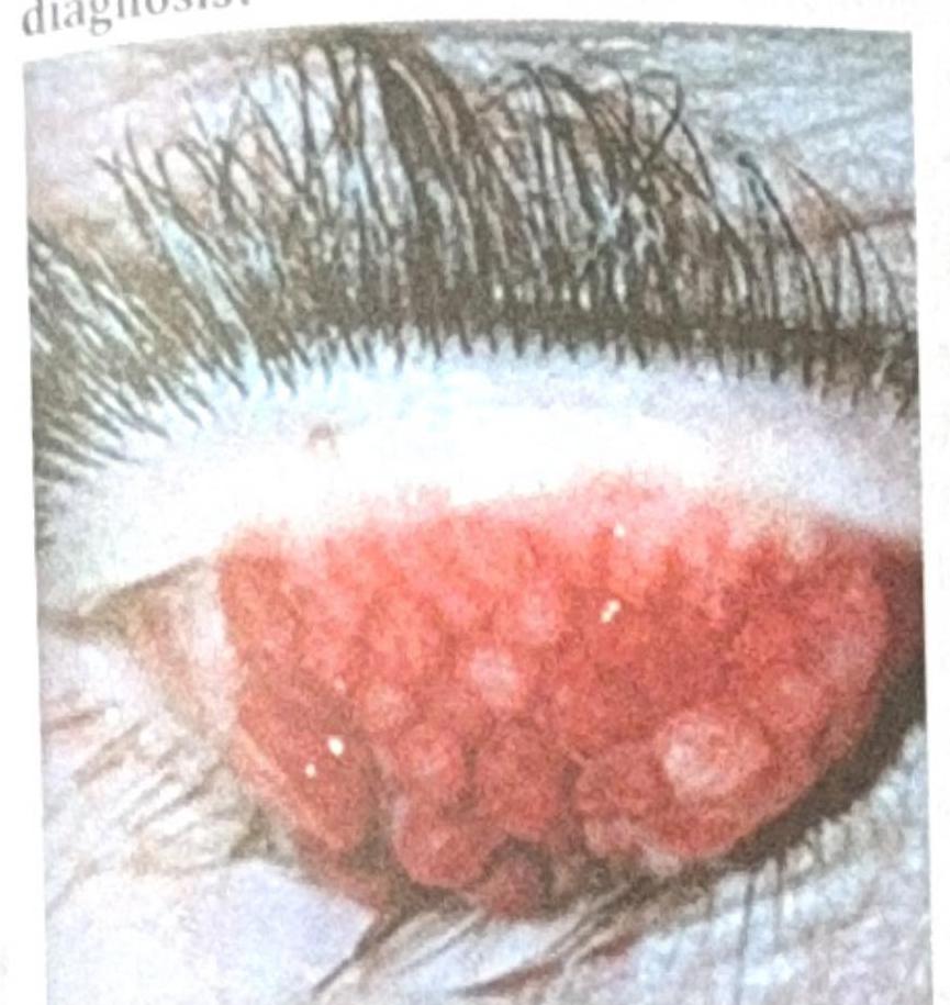
A patient with contact lens use for the past 2 years presents with the ocular findings shown in the image below. What is the most probable diagnosis?

Features of vernal conjunctivitis are:
Epithelial xerosis of conjunctiva is caused by?
NOT a feature of trachoma:
Explanation: ***Staphylococcus*** - **Phlyctenular conjunctivitis** is characterized by delayed (Type IV) hypersensitivity reactions to bacterial antigens, most commonly from **Staphylococcus aureus**. - This condition often presents with small, nodular lesions (phlyctenules) on the conjunctiva or cornea, which are essentially collections of inflammatory cells responding to bacterial proteins. - **Important note**: **Mycobacterium tuberculosis** is another well-documented cause of phlyctenular conjunctivitis, particularly in TB-endemic regions, and should be considered in the differential diagnosis. - Other triggers include protein antigens from organisms colonizing the ocular surface. *Chlamydia* - While **Chlamydia trachomatis** can cause chronic conjunctivitis (e.g., trachoma, adult inclusion conjunctivitis), it does not typically lead to the distinct nodular lesions seen in phlyctenular conjunctivitis. - Ocular chlamydial infections are primarily characterized by follicular conjunctivitis and pannus formation. *Pneumococcus* - **Streptococcus pneumoniae** (Pneumococcus) is a common cause of acute bacterial conjunctivitis, characterized by purulent discharge and redness. - However, it is not associated with the specific delayed hypersensitivity reaction that defines phlyctenular conjunctivitis. *Aspergillus* - **Aspergillus** species are fungi and are more commonly implicated in fungal keratitis or allergic bronchopulmonary aspergillosis, particularly in immunocompromised individuals. - Fungal infections of the conjunctiva are rare and do not typically manifest as phlyctenular conjunctivitis.
Explanation: ***Arise from any part of conjunctiva*** **(FALSE - Correct Answer)** - This statement is **FALSE** and thus the correct answer. - Pterygium characteristically arises from the **nasal (interpalpebral) bulbar conjunctiva** in 90-95% of cases. - It does NOT arise from "any part" - it has a specific predilection for the medial (nasal) limbus in the palpebral fissure zone. - Temporal pterygium is much less common (~10% of cases). *Can cause astigmatism* **(TRUE)** - This statement is TRUE. - As a pterygium grows across the cornea, it can induce **corneal astigmatism** by altering the curvature of the cornea. - This irregular corneal surface can blur vision, especially as the pterygium progresses towards the central visual axis. *Surgery is treatment of choice* **(TRUE)** - This statement is TRUE. - **Surgical excision** is the primary treatment for pterygium when it is symptomatic, threatens vision, or causes significant cosmetic concerns. - Indications for surgery include: growth towards the visual axis, inducing high astigmatism, significant discomfort, or cosmetic desire. - Adjunctive measures (mitomycin C, conjunctival autograft) help reduce recurrence. *UV exposure is risk factor* **(TRUE)** - This statement is TRUE. - **Ultraviolet (UV) radiation exposure** is a well-established and significant risk factor for the development and progression of pterygium. - This explains its higher prevalence in individuals living in sunny climates (between 37° N and 37° S latitude - "pterygium belt") and those with outdoor occupations.
Explanation: **Pterygium** - **Stocker's line** is a **ferrous deposit** that appears as a brown or yellow line at the leading edge of a **pterygium**. - Its presence signifies the progressive nature of the pterygium, indicating ongoing iron deposition due to chronic epithelial degeneration and remodeling. *Glaucoma* - Glaucoma is characterized by **optic nerve damage** and **visual field loss**, usually associated with elevated intraocular pressure. - It does not involve the formation of Stocker's line, which is a corneal or conjunctival finding. *Posterior scleritis* - Posterior scleritis is an **inflammation of the sclera** behind the equator of the globe, often presenting with pain, vision loss, or choroidal folds. - It does not involve the characteristic Stocker's line, which is specific to pterygium. *Diabetic retinopathy* - Diabetic retinopathy involves **microvascular damage** to the retina, leading to vision loss, and is characterized by microaneurysms, hemorrhages, and neovascularization. - It is a retinal disease and does not present with Stocker's line.
Explanation: ***Deleterious effects of foreign bodies*** - This is the **MOST SPECIFIC and PRIMARY reason** that distinguishes retained IOFBs from perforating injuries without retained foreign bodies. - Retained intraocular foreign bodies cause **direct toxic effects** on ocular tissues depending on their composition: **siderosis bulbi** from iron (causing rust-colored deposits, retinal degeneration, and vision loss), **chalcosis** from copper (greenish deposits and inflammation), and direct mechanical trauma to delicate intraocular structures. - These **material-specific toxic effects** are unique to retained foreign bodies and occur regardless of whether infection or inflammation develops. - The foreign body acts as a constant source of **chronic inflammation and tissue damage**, leading to complications like cataract, glaucoma, retinal detachment, and progressive vision loss. *More chances of infection* - While retained IOFBs do increase the risk of **endophthalmitis** (severe intraocular infection), infection risk exists with any perforating injury, whether or not a foreign body is retained. - The question asks what makes retained IOFB cases **MORE serious** - the infection risk is elevated but not the PRIMARY distinguishing feature. - Prophylactic antibiotics can reduce infection risk, but cannot prevent the direct toxic effects of the retained material. *More chances of sympathetic ophthalmitis* - Sympathetic ophthalmitis is a rare bilateral granulomatous uveitis that can occur after **penetrating ocular trauma with uveal tissue injury**. - This risk exists with perforating injuries in general, not specifically because of the retained foreign body itself. - The presence of a foreign body is less important than uveal prolapse and inflammation in triggering this immune-mediated response. *All of the options* - While infection and sympathetic ophthalmitis are legitimate concerns, they are **not specific to retained foreign bodies** - they can occur with any penetrating injury. - The **direct deleterious/toxic effects** of the foreign body material (siderosis, chalcosis, mechanical damage) are the PRIMARY and MOST SPECIFIC reason that makes retained IOFB cases more serious. - This option is incorrect because it doesn't distinguish the unique hazard posed by the retained foreign body itself.
Explanation: ***Correct: Statements 1 & 2 are correct, 2 is not explaining 1*** **Analysis of Statement 1:** - A 59-year-old patient with **flaccid bullae** and **suprabasal acantholytic split** on histopathology is the classic presentation of **Pemphigus vulgaris** - The flaccid (easily ruptured) nature of bullae distinguishes it from tense bullae seen in bullous pemphigoid - The suprabasal location of the split (just above the basal layer) with acantholysis (loss of cell-to-cell adhesion) is pathognomonic - **Statement 1 is CORRECT** ✓ **Analysis of Statement 2:** - The **"row of tombstones" or "tombstone appearance"** is indeed a diagnostic histopathological feature of Pemphigus vulgaris - This appearance results from basal keratinocytes remaining attached to the basement membrane while suprabasal cells separate due to acantholysis - The intact basal cells standing upright resemble a row of tombstones - **Statement 2 is CORRECT** ✓ **Does Statement 2 explain Statement 1?** - Statement 2 describes a **histopathological appearance** (tombstone pattern) that is a **consequence** of the suprabasal split - However, it does NOT explain the **underlying cause** of the flaccid bullae or the suprabasal split - The true explanation involves **IgG autoantibodies against desmoglein 3 (and desmoglein 1)**, which attack intercellular adhesion structures (desmosomes), causing **acantholysis** - Therefore, **Statement 2 does NOT explain Statement 1** ✗ *Incorrect: Statement 2 is the correct explanation for Statement 1* - While both statements describe features of Pemphigus vulgaris, the tombstone appearance is a descriptive finding, not an explanatory mechanism *Incorrect: Statements 1 and 2 are incorrect* - Both statements are medically accurate descriptions of Pemphigus vulgaris features *Incorrect: Statement 1 is incorrect* - Statement 1 correctly describes the cardinal clinical and histopathological features of Pemphigus vulgaris
Explanation: ***Iris*** - **Koeppe nodules** are characteristic inflammatory granulomas found along the **pupillary margin** of the iris. - They are typically seen in **granulomatous uveitis**, particularly in conditions like sarcoidosis or tuberculosis. *Cornea* - The cornea is the transparent front part of the eye that covers the iris, pupil, and anterior chamber. - While corneal changes like **keratic precipitates** can occur in uveitis, **Koeppe nodules** do not develop on the cornea itself. *Conjunctiva* - The conjunctiva is the mucous membrane lining the inner surface of the eyelids and covering the anterior sclera. - Inflammatory conditions of the conjunctiva may present with follicles or papillae, but **Koeppe nodules** are not found here. *Retina* - The retina is the light-sensitive tissue at the back of the eye, responsible for vision. - Retinal involvement in uveitis can lead to vasculitis or macula edema, but **Koeppe nodules** are specific to the iris.
Explanation: ***Giant Papillary conjunctivitis*** - The image shows **large, elevated papillae** on the **tarsal conjunctiva**, which are characteristic findings of giant papillary conjunctivitis. - This condition is common among **contact lens wearers**, caused by chronic mechanical irritation and an allergic response to lens material or deposits. *Trachoma* - Trachoma is a **chronic infectious disease** caused by *Chlamydia trachomatis*, leading to scarring of the conjunctiva. - It typically presents with **follicles** in the early stages, followed by **scarring** and **pannus formation**, not the large papillae seen here. *Ocular Surface Squamous Neoplasia (OSSN)* - OSSN refers to a spectrum of conditions from **dysplasia to squamous cell carcinoma** affecting the conjunctiva or cornea. - It usually presents as a **gelatinous, fleshy, or leukoplakic lesion**, often at the limbus, which is distinct from the diffuse papillae shown. *Vernal Keratoconjunctivitis* - Vernal keratoconjunctivitis (VKC) is a **severe form of allergic conjunctivitis** but primarily affects children and young adults with a history of atopy. - While it can cause large papillae (cobblestone papillae), it is not specifically associated with contact lens wear and usually has other systemic allergic manifestations.
Explanation: ***All of the options*** - **Vernal conjunctivitis (VKC)** is a severe form of allergic conjunctivitis characterized by chronic inflammation of the conjunctiva, impacting the cornea in advanced stages. - **Shield ulcers**, **Horner-Trantas dots**, and **papillary hypertrophy** are all classic clinical features observed in VKC. *Shield ulcer* - This is a **corneal complication** of severe vernal conjunctivitis, characterized by epithelial defects that can lead to significant pain and vision impairment. - It develops due to corneal abrasion from the giant papillae on the upper tarsal conjunctiva and direct corneal toxicity from inflammatory mediators. *Horner-Trantas spots* - These are **gelatinous aggregations** of epithelial cells and eosinophils that appear as white dots at the limbus, particularly evident at the superior limbus. - They are one of the **pathognomonic signs** of vernal conjunctivitis, indicating significant allergic inflammation. *Papillary hypertrophy* - Characterized by the development of **large, flattened papillae** (often described as "cobblestone" papillae) on the upper tarsal conjunctiva. - This hypertrophy is a result of chronic inflammation and proliferation of mast cells, eosinophils, and lymphocytes in the conjunctival stroma.
Explanation: ***Xerophthalmia*** - **Xerophthalmia** is a medical condition characterized by **dryness of the eye**, often due to **vitamin A deficiency**. - **Epithelial xerosis of the conjunctiva** is one of the early and hallmark signs of xerophthalmia, representing the drying and thickening of the conjunctival epithelium due to goblet cell loss and squamous metaplasia. *Infectious conjunctivitis caused by Chlamydia trachomatis* - This typically causes **trachoma**, characterized by chronic inflammation, scarring, and eventual blindness. - While it can lead to dryness and scarring in later stages due to **symblepharon** or **entropion**, it does not primarily manifest as epithelial xerosis. *Autoimmune blistering conjunctivitis* - This condition involves **immune-mediated inflammation** leading to subepithelial blistering, scarring, and shrinkage of the conjunctiva. - It results in significant **ocular surface damage** and vision loss but is distinct from the primary epithelial changes seen in xerosis due to vitamin A deficiency. *Bacterial conjunctivitis due to Corynebacterium diphtheriae* - **Diphtheritic conjunctivitis** is a severe form of bacterial conjunctivitis that causes a distinctive **"pseudomembrane"** on the conjunctiva. - It leads to acute inflammation and potentially systemic illness, not primarily epithelial xerosis.
Explanation: ***Chalazion*** - A **chalazion** is a **lipogranulomatous inflammation** of a **meibomian gland** and is not directly caused by *Chlamydia trachomatis* infection, though chronic inflammation could theoretically predispose to it. - While chronic inflammation of the eyelids in trachoma can cause various complications, a chalazion is a distinct condition related to meibomian gland dysfunction and is not a direct, defining feature of trachoma. *Entropion* - **Entropion**, the **inward turning of the eyelid margin**, is a severe late complication of trachoma caused by conjunctival scarring and contraction. - This inward turning leads to **trichiasis** (**misdirected eyelashes**), which abrades the cornea. *Corneal opacity* - **Corneal opacity** is a common and serious consequence of chronic trachoma, resulting from repeated **corneal abrasions** by misdirected eyelashes (trichiasis) and chronic inflammation. - This scarring can lead to **severe vision impairment** and **blindness**. *Herbert's pits* - **Herbert's pits** are characteristic depressions on the **limbus** (corneoscleral junction) formed after the resolution of **limbal follicles** in chronic trachoma. - They are a diagnostic sign of past or present trachomatous infection.
Get full access to all questions, explanations, and performance tracking.
Start For Free