Which of the following findings is typically NOT seen in a patient with allergic conjunctivitis?
A 25-year-old male presents with recurrent bilateral conjunctival hyperemia and a gritty sensation. Likely diagnosis?
Arlt's line is seen in?
Phlyctenular conjunctivitis is primarily associated with hypersensitivity to which of the following?
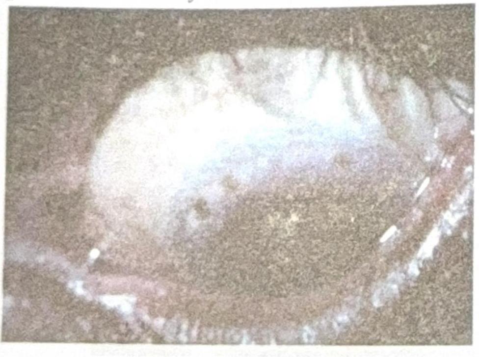
Identify the diagnosis based on the clinical image shown.

Which of the following is not a treatment option for vernal keratoconjunctivitis?
Inclusion conjunctivitis is caused by:
Features of vernal conjunctivitis are:
Unilateral frontal blisters with upper lid edema and conjunctivitis is seen in?
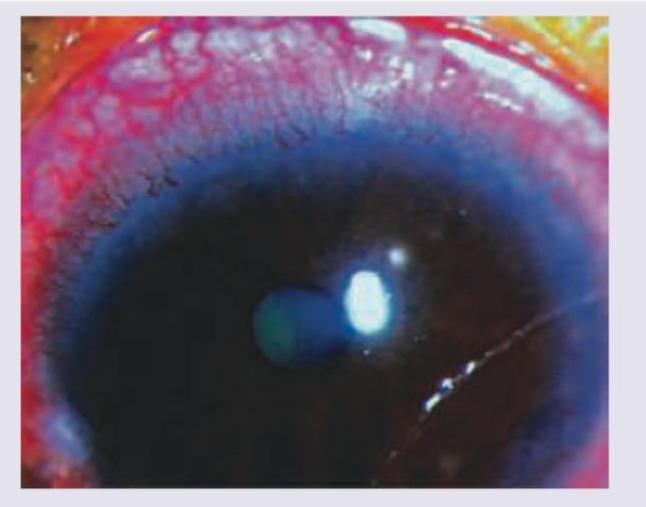
What does the following image show?

Explanation: ***Purulent discharge*** - **Purulent discharge** (thick, yellowish, or greenish) is characteristic of **bacterial conjunctivitis** and is usually absent in allergic conjunctivitis. - Allergic conjunctivitis typically presents with a **clear or watery discharge**. *Watery discharge* - **Watery discharge** is a very common symptom of **allergic conjunctivitis**, often accompanied by itching and redness. - It results from the inflammatory response and increased lacrimation due to allergen exposure. *Itching* - **Ocular itching** is the hallmark symptom of allergic conjunctivitis and is considered its most distinctive feature. - It is caused by the release of **histamine** and other inflammatory mediators from mast cells in response to allergens. *Bilateral eye redness* - **Bilateral conjunctival redness** (hyperemia) is a frequent finding in allergic conjunctivitis. - This is due to **vasodilation** in response to the inflammatory process affecting both eyes, as airborne allergens often affect both simultaneously.
Explanation: ***Vernal keratoconjunctivitis*** * This is the correct diagnosis as it perfectly matches the clinical presentation: **young male patient** (VKC has male predominance, especially in adolescents/young adults), **recurrent course** (VKC is a chronic allergic condition with seasonal exacerbations), and **bilateral involvement** with gritty sensation. * VKC is a **severe form of allergic conjunctivitis** characterized by **bilateral conjunctival hyperemia**, intense itching, gritty sensation, photophobia, and mucoid discharge. The recurrent bilateral nature in a young male is pathognomonic. *Herpes keratitis* * Usually presents as **unilateral eye pain**, redness, and a characteristic **dendritic ulcer** on the cornea (seen with fluorescein staining), which is not described here. * Caused by herpes simplex virus and typically has an acute presentation rather than recurrent bilateral conjunctival symptoms. Can lead to significant vision loss if untreated. *Episcleritis* * Characterized by **localized sectorial redness** in one eye, often in a radial pattern, and is usually **mild and self-limiting**. * Typically causes minimal discomfort and does not commonly present with gritty sensation or recurrent bilateral involvement as the primary feature. *Bacterial conjunctivitis* * Typically presents with **purulent discharge** (thick yellow-green pus) and matting of eyelids, which is not mentioned in this patient's symptoms. * While it causes redness and grittiness, it's usually **acute and unilateral or sequential bilateral** (one eye then the other), and resolves with topical antibiotics within days, unlike the recurrent chronic nature described here.
Explanation: ***Trachoma*** - Arlt's line is a **subtarsal fibrous white line** seen on the upper tarsal conjunctiva, resulting from repeated episodes of inflammation and scarring in **trachoma**. - This scarring can lead to **entropion** and **trichiasis**, causing corneal abrasion and potential blindness. *Vernal catarrh* - Characterized by giant papillae (cobblestone papillae) on the upper tarsal conjunctiva and **Horner-Trantas dots** at the limbus. - It is an allergic condition, and while it causes conjunctival changes, it does not typically result in Arlt's line. *Allergic conjunctivitis* - Presents with itching, redness, tearing, and sometimes mild conjunctival swelling or papillae. - It is an acute or chronic allergic reaction and does not cause the specific scarring pattern known as Arlt's line. *Bacterial conjunctivitis* - Typically presents with **purulent discharge**, redness, and lid crusting. - While it causes acute inflammation, it usually resolves without the chronic scarring that leads to Arlt's line unless it is a severe, recurrent infection.
Explanation: ***Staphylococcus*** - **Phlyctenular conjunctivitis** is characterized by delayed (Type IV) hypersensitivity reactions to bacterial antigens, most commonly from **Staphylococcus aureus**. - This condition often presents with small, nodular lesions (phlyctenules) on the conjunctiva or cornea, which are essentially collections of inflammatory cells responding to bacterial proteins. - **Important note**: **Mycobacterium tuberculosis** is another well-documented cause of phlyctenular conjunctivitis, particularly in TB-endemic regions, and should be considered in the differential diagnosis. - Other triggers include protein antigens from organisms colonizing the ocular surface. *Chlamydia* - While **Chlamydia trachomatis** can cause chronic conjunctivitis (e.g., trachoma, adult inclusion conjunctivitis), it does not typically lead to the distinct nodular lesions seen in phlyctenular conjunctivitis. - Ocular chlamydial infections are primarily characterized by follicular conjunctivitis and pannus formation. *Pneumococcus* - **Streptococcus pneumoniae** (Pneumococcus) is a common cause of acute bacterial conjunctivitis, characterized by purulent discharge and redness. - However, it is not associated with the specific delayed hypersensitivity reaction that defines phlyctenular conjunctivitis. *Aspergillus* - **Aspergillus** species are fungi and are more commonly implicated in fungal keratitis or allergic bronchopulmonary aspergillosis, particularly in immunocompromised individuals. - Fungal infections of the conjunctiva are rare and do not typically manifest as phlyctenular conjunctivitis.
Explanation: ***Herbert's pits*** - The image displays characteristic **pits at the limbus**, which are a hallmark of healed follicular conjunctivitis, specifically **Herbert's pits**. - These pits are pathognomonic for **trachoma**, a chronic keratoconjunctivitis caused by *Chlamydia trachomatis*. *Horner-Trantas spots* - These are small, raised, gelatinous white dots found at the limbus, typically containing **eosinophils**. - They are characteristic of **vernal keratoconjunctivitis**, an allergic inflammatory condition, and not seen in the provided image. *Pannus* - **Pannus** refers to the growth of **vascularized connective tissue** onto the cornea. - While pannus is also a feature of trachoma, the image specifically shows distinct limbal pits, rather than diffuse vascularization. *Corneal dystrophy* - **Corneal dystrophies** are a group of inherited, bilateral, and progressive corneal disorders that manifest as opacities or structural changes in the cornea. - The findings in the image, such as pits, are inflammatory in origin and distinct from the typically stromal or epithelial changes seen in corneal dystrophies.
Explanation: ***Antibiotics*** - **Vernal keratoconjunctivitis (VKC)** is a non-infectious, **allergic inflammatory condition** of the conjunctiva. - Antibiotics are primarily used to treat bacterial infections and have **no direct role** in the management of VKC. *Steroids* - **Topical corticosteroids** (e.g., prednisolone, loteprednol, fluorometholone) are a mainstay of VKC treatment, especially for severe cases and acute exacerbations. - They significantly reduce inflammation and associated symptoms by **suppressing the immune response**. *Chromaglycate* - **Sodium cromoglycate** is a **mast cell stabilizer** commonly used in the treatment of VKC. - It works by preventing the degranulation of mast cells, thereby **inhibiting the release of inflammatory mediators** like histamine. *Olopatadine* - **Olopatadine** is a dual-acting medication that functions as both an **antihistamine** and a **mast cell stabilizer**. - It provides rapid relief from itching and other allergic symptoms by blocking histamine receptors and stabilizing mast cells.
Explanation: ***Chlamydia trachomatis*** - **Inclusion conjunctivitis** is primarily caused by **Chlamydia trachomatis serovars D-K**. - This form of conjunctivitis is characterized by follicular response and cytoplasmic inclusions seen in epithelial cells. *Klebsiella species* - **Klebsiella** are common bacteria that can cause a variety of infections, including pneumonia and urinary tract infections. - While they can cause conjunctivitis, it is typically a bacterial conjunctivitis and not specifically referred to as inclusion conjunctivitis. *Adenovirus* - **Adenoviruses** are a common cause of **viral conjunctivitis**, which often presents with watery discharge and pharyngitis. - Unlike *Chlamydia*, adenovirus infections do not form characteristic cytoplasmic inclusions in conjunctival cells. *Mycobacterium Leprae* - **Mycobacterium leprae** is the causative agent of **Leprosy**, a chronic infectious disease primarily affecting the skin, nerves, and upper respiratory tract. - It does not directly cause conjunctivitis as its primary manifestation, although ocular complications can occur in advanced leprosy.
Explanation: ***All of the options*** - **Vernal conjunctivitis (VKC)** is a severe form of allergic conjunctivitis characterized by chronic inflammation of the conjunctiva, impacting the cornea in advanced stages. - **Shield ulcers**, **Horner-Trantas dots**, and **papillary hypertrophy** are all classic clinical features observed in VKC. *Shield ulcer* - This is a **corneal complication** of severe vernal conjunctivitis, characterized by epithelial defects that can lead to significant pain and vision impairment. - It develops due to corneal abrasion from the giant papillae on the upper tarsal conjunctiva and direct corneal toxicity from inflammatory mediators. *Horner-Trantas spots* - These are **gelatinous aggregations** of epithelial cells and eosinophils that appear as white dots at the limbus, particularly evident at the superior limbus. - They are one of the **pathognomonic signs** of vernal conjunctivitis, indicating significant allergic inflammation. *Papillary hypertrophy* - Characterized by the development of **large, flattened papillae** (often described as "cobblestone" papillae) on the upper tarsal conjunctiva. - This hypertrophy is a result of chronic inflammation and proliferation of mast cells, eosinophils, and lymphocytes in the conjunctival stroma.
Explanation: ***Herpes Zoster Ophthalmicus*** - This condition is characterized by a **unilateral vesicular rash** (blisters) in the **trigeminal dermatome (V1)**, which includes the forehead and upper eyelid, along with significant **lid edema** and **conjunctivitis**. - **Hutchinson's sign** (lesions on the tip, side, or root of the nose) indicates a high risk of ocular involvement due to the nasociliary nerve innervation. *Acanthamoeba Keratitis* - This is an **amoebic infection** of the cornea typically associated with **contact lens wear** and often presents with severe pain and a **ring infiltrate** in the cornea. - It does not typically present with unilateral frontal blisters or significant lid edema. *Herpes Simplex* - Herpes simplex typically causes **recurrent corneal ulcers** (dendritic or geographic) and sometimes blepharitis, but not the widespread **unilateral frontal blisters** seen in the trigeminal distribution. - While it can cause conjunctivitis and lid edema, the pattern of skin lesions is the key differentiator. *Neuroparalytic Keratitis* - This condition results from **trigeminal nerve damage**, leading to corneal anesthesia and subsequent **trophic corneal ulceration**. - It presents primarily with **corneal findings** (epithelial defects, ulcers) due to impaired sensation and tear film stability, not initial vesicular skin lesions or prominent lid edema.
Explanation: ***Pannus*** - The image distinctly shows **new blood vessels growing into the cornea** from the limbus, which is characteristic of pannus formation. This vascularization often accompanies chronic inflammation or hypoxia. - This condition is a hallmark of certain ophthalmic diseases, such as **trachoma** or chronic irritation from prolonged **contact lens wear**. *Anterior uveitis* - Anterior uveitis presents with inflammation of the iris and ciliary body, typically causing **ciliary flush**, **miosis**, and cells/flare in the anterior chamber, which are not the primary features shown. - While uveitis can sometimes lead to corneal changes, the prominent **vascularization extending onto the cornea** is not its defining visual characteristic. *Membranous conjunctivitis* - Membranous conjunctivitis is characterized by the formation of a **true membrane on the conjunctiva** that binds tightly to the underlying epithelium, often caused by severe bacterial infections like *Corynebacterium diphtheriae*. - The image does not show a membrane on the conjunctival surface; instead, it highlights **corneal vascularization**. *Pseudomembranous conjunctivitis* - Pseudomembranous conjunctivitis involves a **fibrinous exudate loosely adherent** to the conjunctiva, which can be peeled off without significant bleeding, as seen in adenoviral conjunctivitis. - The findings in the image, specifically **vascular ingrowth into the cornea**, are not consistent with the appearance of a pseudomembrane on the conjunctiva.
Get full access to all questions, explanations, and performance tracking.
Start For Free