Recovery in cataract surgery is fastest with which of the following techniques?
What is the equatorial diameter of the lens?
In which of the following uveitic conditions is it contraindicated to implant an intraocular lens after cataract extraction?
What is the best method to prevent infection after cataract surgery?
What causes a 'Rosette cataract'?
Lens develops from which germ layer?
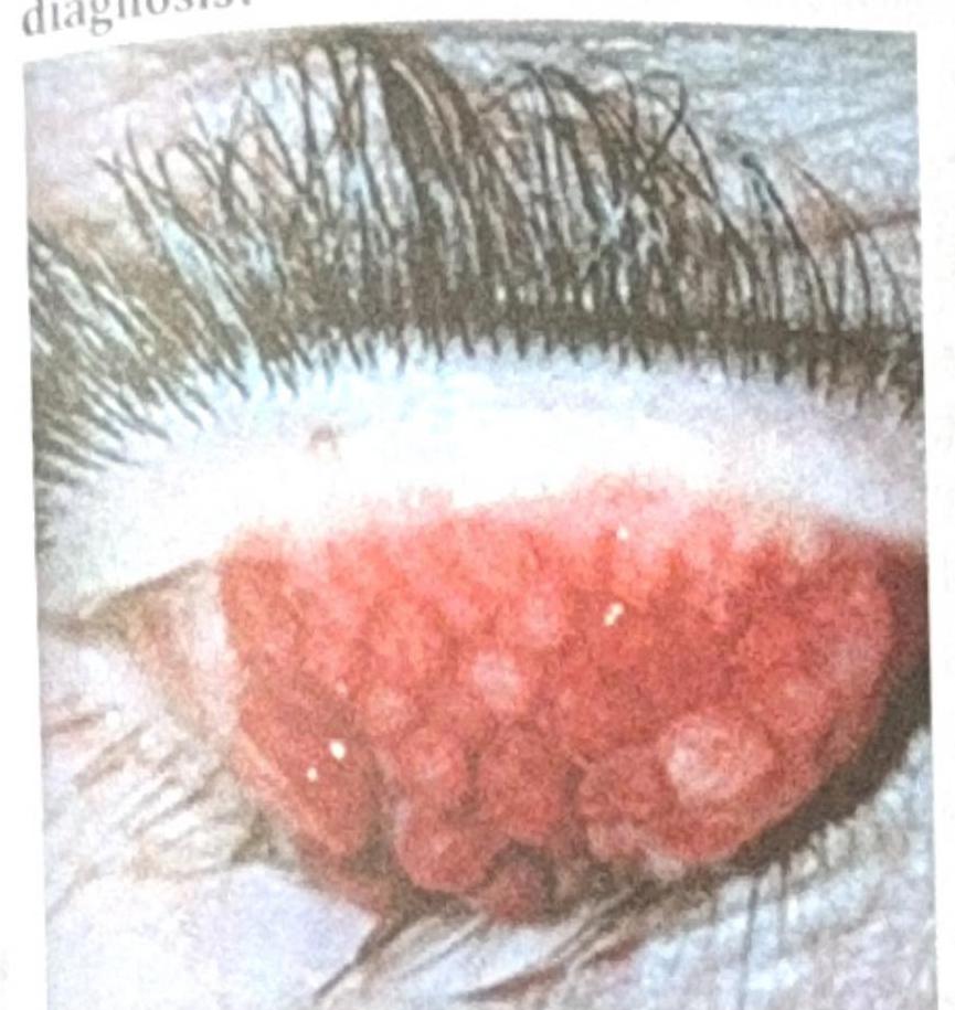
A patient with contact lens use for the past 2 years presents with the ocular findings shown in the image below. What is the most probable diagnosis?

Which of the following is an advantage of contact lenses over normal glasses?
Rigid gas permeable (RGP) lenses are made from which of the following combinations of materials?
What is the most common infection in contact lens users?
Explanation: **Explanation:** The speed of visual and physical recovery after cataract surgery is primarily determined by the **size of the incision** and the resulting **surgically induced astigmatism (SIA)**. **Why Phacoemulsification is correct:** Phacoemulsification is a "small-incision" surgery (typically 2.2 to 2.8 mm). Because the incision is so small, it is often **self-sealing (sutureless)**. This leads to: * **Minimal Astigmatism:** Rapid stabilization of the corneal curvature. * **Early Visual Rehabilitation:** Patients often achieve near-optimal vision within days. * **Structural Integrity:** The eye regains its strength almost immediately, allowing for a quicker return to physical activities. **Why other options are incorrect:** * **ICCE:** This involves a large 120–140 degree superior limbal incision (approx. 10–12 mm). It requires multiple sutures, leads to high astigmatism, and has a high risk of vitreous loss. Recovery takes months. * **ECCE:** Traditional ECCE requires a 10–12 mm incision to deliver the nucleus in one piece. The large wound requires sutures, leading to significant SIA and a recovery period of 6–8 weeks. * **ECCE with IOL:** While this improves long-term visual quality compared to aphakia, the recovery speed remains slow due to the large incision size required for the procedure. **High-Yield Clinical Pearls for NEET-PG:** * **Gold Standard:** Phacoemulsification is the current gold standard for cataract surgery. * **SICS (Manual Small Incision Cataract Surgery):** Uses a 6–7 mm valvular tunnel. It is faster than ECCE but slower than Phacoemulsification in terms of visual stabilization. * **Astigmatism:** Large incisions (ECCE/ICCE) typically cause **"With-the-rule"** astigmatism initially, which may shift over time. Small incisions in Phacoemulsification are considered "astigmatically neutral."
Explanation: ### Explanation The human crystalline lens is a transparent, biconvex structure located behind the iris. Understanding its dimensions is crucial for NEET-PG, as these parameters change with age and accommodation. **Why 9 mm is correct:** At birth, the equatorial diameter of the lens is approximately **6.5 mm**. However, as the lens grows throughout life, it reaches an average adult diameter of **9 to 10 mm**. In the context of standard medical examinations and textbooks (like AK Khurana), **9 mm** is the most frequently cited and accepted value for the adult equatorial diameter. **Analysis of Incorrect Options:** * **A (7 mm):** This is closer to the diameter at birth (6.5 mm). It is too small for a fully developed adult lens. * **B (8 mm):** This represents an intermediate stage of development and is not the standard adult measurement. * **D (10 mm):** While the lens can reach 10 mm in older age, 9 mm is the classic "textbook" answer for the adult lens diameter. **High-Yield Clinical Pearls for NEET-PG:** * **Thickness:** The anteroposterior thickness of the lens is approximately **3.5 to 4 mm** at birth, increasing to about **4.5 to 5 mm** in old age. * **Radius of Curvature:** The anterior surface is flatter (radius: **10 mm**), while the posterior surface is more curved (radius: **6 mm**). * **Refractive Power:** The lens contributes approximately **15–18 Diopters** to the total refractive power of the eye (which is ~60D). * **Refractive Index:** The average refractive index of the lens is **1.39**, but it varies from the cortex (1.38) to the nucleus (1.41)—this is known as the *gradient refractive index*. * **Composition:** The lens has the highest protein content of any tissue in the body (about 33%).
Explanation: **Explanation:** The correct answer is **Juvenile Rheumatoid Arthritis (JRA)**, specifically the pauciarticular type associated with chronic non-granulomatous uveitis. **1. Why JRA is the correct answer:** In JRA-associated uveitis, the eye is prone to chronic, recalcitrant inflammation. Implanting an Intraocular Lens (IOL) in these patients is traditionally contraindicated (or approached with extreme caution) because it acts as a foreign body scaffold for inflammatory membranes. This leads to a high incidence of **cyclitic membranes**, persistent hypotony, and eventual phthisis bulbi. Most surgeons prefer leaving the patient **aphakic** and managing vision with contact lenses or spectacles once the inflammation is controlled. **2. Why the other options are incorrect:** * **A. Fuchs Heterochromic Cyclitis:** This is a unique form of chronic uveitis where IOL implantation is actually **very successful**. Despite the presence of stellate KPs and iris atrophy, these eyes do not develop posterior synechiae and tolerate IOLs remarkably well. * **C & D. Psoriatic Arthritis and Reiter’s Syndrome:** These are HLA-B27 associated spondyloarthropathies. While they cause acute anterior uveitis, the inflammation is usually episodic rather than chronic. Once the acute attack is controlled, IOL implantation is generally safe and successful. **Clinical Pearls for NEET-PG:** * **Gold Standard for JRA Cataract:** Lensectomy + Anterior Vitrectomy (leaving the eye aphakic). * **Fuchs Heterochromic Cyclitis:** Characterized by the triad of heterochromia, cataract, and glaucoma; it is the "exception" where steroids are generally not required post-operatively. * **Absolute Contraindications for IOL (Traditional):** JRA-associated uveitis and Sympathetic Ophthalmia (though modern surgical techniques are cautiously challenging these).
Explanation: **Explanation:** The primary goal in preventing post-cataract surgery infection (endophthalmitis) is the reduction of microbial load on the ocular surface and the prevention of bacterial entry into the eye. **Why Antibiotic Administration is Correct:** The use of antibiotics is the most evidence-based method for preventing endophthalmitis. Specifically, the **intracameral injection of Cefuroxime** (as demonstrated by the ESCRS study) at the end of surgery significantly reduces the risk of infection. Additionally, preoperative application of **Povidone-Iodine (5%)** to the conjunctival sac is considered the "gold standard" for antisepsis, as it kills bacteria more effectively than topical antibiotics alone. **Why Other Options are Incorrect:** * **Eyebrow Shaving:** This is an outdated practice. Shaving can cause micro-abrasions on the skin, which act as a nidus for bacterial colonization and actually *increase* the risk of surgical site infections. * **Irrigation of the Surgical Site:** While irrigation (hydrodissection/aspiration) is a standard part of the surgical procedure to remove lens matter, it is not a primary prophylactic measure against infection. In fact, excessive irrigation without proper aseptic technique can theoretically introduce contaminants. **Clinical Pearls for NEET-PG:** 1. **Most common organism** causing acute post-operative endophthalmitis: *Staphylococcus epidermidis* (Coagulase-negative Staph). 2. **Most common source** of infection: The patient’s own conjunctival and eyelid flora. 3. **Prophylaxis Gold Standard:** 5% Povidone-Iodine (Betadine) in the conjunctival sac (left for 3 minutes) and 10% on the periocular skin. 4. **Intracameral Cefuroxime (1mg in 0.1ml)** is the most effective pharmacological intervention to prevent endophthalmitis post-Phacoemulsification.
Explanation: **Explanation:** **Correct Answer: C. Trauma** A **Rosette-shaped cataract** is a classic sign of blunt ocular trauma. When the eye sustains a mechanical impact, hydraulic shockwaves travel through the lens. This leads to the separation of lens fibers along their natural suture lines, particularly in the posterior subcapsular cortex. The resulting opacification follows the anatomical pattern of the lens sutures, creating a characteristic "flower-shaped" or "star-shaped" appearance. While it typically appears shortly after trauma, it can sometimes remain stationary or progress over years. **Analysis of Incorrect Options:** * **A. Diabetes:** Diabetic cataracts typically present as **"Snowflake cataracts"** (subcapsular opacities) due to osmotic swelling caused by sorbitol accumulation. * **B. After cataract:** Also known as Posterior Capsular Opacification (PCO), this occurs following cataract surgery. Common forms include **Elschnig’s pearls** or **Soemmering’s ring**, but not rosette patterns. * **D. Gaucher's disease:** This lysosomal storage disorder is not typically associated with rosette cataracts; it more commonly presents with pinguecula-like lesions or retinal findings. **High-Yield Clinical Pearls for NEET-PG:** * **Vossius Ring:** A circular ring of pigment on the anterior lens capsule, also caused by blunt trauma (imprint of the iris). * **Sunflower Cataract:** Associated with **Wilson’s Disease** (copper deposition). * **Oil Droplet Cataract:** Pathognomonic for **Galactosemia**. * **Christmas Tree Cataract:** Seen in **Myotonic Dystrophy**. * **Shield Cataract:** Associated with **Atopic Dermatitis**.
Explanation: **Explanation:** The development of the eye is a complex process involving multiple germ layers. The **Surface Ectoderm** is the correct answer because the lens originates from the **lens placode**, a thickening of the surface ectoderm that occurs when the underlying optic vesicle (from the neuroectoderm) comes into contact with it. This placode eventually invaginates to form the lens vesicle, which detaches to become the crystalline lens. **Analysis of Options:** * **Surface Ectoderm (Correct):** Besides the **lens**, it gives rise to the corneal epithelium, conjunctival epithelium, lacrimal apparatus, and the epithelium of the eyelids/eyelashes. * **Neuroectoderm (Incorrect):** This layer forms the "neural" components of the eye, including the **retina**, posterior layers of the iris, ciliary body epithelium, and the **optic nerve**. * **Mesoderm (Incorrect):** It contributes primarily to the extraocular muscles, vascular endothelium, and the temporal portion of the sclera. * **Endoderm (Incorrect):** The endoderm does not contribute to the formation of any ocular structures. **High-Yield Clinical Pearls for NEET-PG:** * **Neural Crest Cells:** These are vital for the "structural" framework. They form the **corneal stroma/endothelium**, most of the **sclera**, and the trabecular meshwork. * **Lens Nutrition:** In fetal life, the lens is nourished by the **hyaloid artery** (tunica vasculosa lentis). Failure of this system to regress leads to Persistent Hyperplastic Primary Vitreous (PHPV). * **Aphakia:** The absence of the lens (congenital or surgical) results in a high degree of hypermetropia.
Explanation: ***Giant Papillary conjunctivitis*** - The image shows **large, elevated papillae** on the **tarsal conjunctiva**, which are characteristic findings of giant papillary conjunctivitis. - This condition is common among **contact lens wearers**, caused by chronic mechanical irritation and an allergic response to lens material or deposits. *Trachoma* - Trachoma is a **chronic infectious disease** caused by *Chlamydia trachomatis*, leading to scarring of the conjunctiva. - It typically presents with **follicles** in the early stages, followed by **scarring** and **pannus formation**, not the large papillae seen here. *Ocular Surface Squamous Neoplasia (OSSN)* - OSSN refers to a spectrum of conditions from **dysplasia to squamous cell carcinoma** affecting the conjunctiva or cornea. - It usually presents as a **gelatinous, fleshy, or leukoplakic lesion**, often at the limbus, which is distinct from the diffuse papillae shown. *Vernal Keratoconjunctivitis* - Vernal keratoconjunctivitis (VKC) is a **severe form of allergic conjunctivitis** but primarily affects children and young adults with a history of atopy. - While it can cause large papillae (cobblestone papillae), it is not specifically associated with contact lens wear and usually has other systemic allergic manifestations.
Explanation: ***Improved peripheral vision*** - Contact lenses sit directly on the cornea, moving with the eye and eliminating the **frame obstruction** and **edge distortions** associated with glasses. - This provides a wider and more natural **field of view**, enhancing peripheral vision. *Reduced prismatic effect* - While contact lenses do reduce the **magnification/minification** compared to glasses, the prismatic effect is a specific distortion most pronounced in **strong thick spectacle lenses** and can induce visual discomfort, which contact lenses inherently minimize. - This effect is due to the distance between the spectacle lens and the eye, which contact lenses eliminate. *Decreased risk of infection* - Wearing contact lenses inherently carries a **higher risk of eye infections** if proper hygiene and care are not meticulously followed. - Unlike glasses, contact lenses require regular cleaning, disinfection, and proper storage to prevent bacterial or fungal contamination. *UV protection (in specific lenses)* - While some contact lenses incorporate **UV-blocking agents**, this is not a universal feature of all contact lenses and is also available in many spectacle lenses. - UV protection from contact lenses primarily shields the cornea and iris but does not fully protect the surrounding ocular tissues like glasses (especially wrap-around styles) can.
Explanation: ***Copolymer of PMMA, Silicon containing monomer & cellulose acetyl butyrate*** - **Rigid gas permeable (RGP) lenses** are designed to be permeable to oxygen, which is achieved through the incorporation of **silicon-containing monomers**. - The combination of **PMMA** (for rigidity), **silicon** (for oxygen permeability), and **cellulose acetyl butyrate** (for improved wettability and flexibility) provides the desired mechanical and optical properties. *Polymethylmethacrylate* - **PMMA** was the primary material for the earliest **hard contact lenses** but offered virtually no oxygen permeability. - This lack of oxygen permeability led to significant corneal hypoxia issues and limited wear time. *Hydroxymethylmethacrylate* - **Hydroxymethylmethacrylate (HEMA)** is a key material in **hydrogel soft contact lenses**, known for its ability to absorb water. - HEMA is not used in RGP lenses because it would make the lens soft and flexible, contrary to the "rigid" characteristic. *Cellulose acetate Butyrate* - **Cellulose acetate butyrate (CAB)** was an early material used for **gas permeable lenses**, offering some oxygen permeability. - While it was an improvement over PMMA, it did not achieve the high level of oxygen permeability seen with newer silicon-containing materials.
Explanation: ***Pseudomonas*** - **Pseudomonas aeruginosa** is the leading cause of **bacterial keratitis** in contact lens wearers, accounting for 60-70% of culture-positive cases - This bacterium can **adhere to lenses**, form **biofilms**, and thrive in moist lens storage cases - Can cause rapid and severe corneal damage with **corneal ulceration**, potentially leading to **vision loss** *Staphylococcus* - **Staphylococcus aureus** and **Staphylococcus epidermidis** are common commensals of the skin and can cause eye infections, including keratitis and blepharitis - However, in the context of contact lens-related keratitis, **Pseudomonas aeruginosa** remains the primary pathogen for severe corneal infections *Streptococcus* - While various **Streptococcus species** (especially S. pneumoniae) can cause bacterial keratitis, they are less commonly associated with contact lens-related keratitis compared to Pseudomonas - **Streptococcal keratitis** typically occurs in non-contact lens wearers or after trauma *Neisseria* - **Neisseria gonorrhoeae** can cause hyperacute bacterial conjunctivitis with severe purulent discharge, but is not the most common cause of contact lens-related keratitis - **Neisseria meningitidis** can rarely cause conjunctivitis, but these infections usually indicate specific exposure or systemic disease
Contact Lens Materials
Practice Questions
Soft Contact Lenses
Practice Questions
Rigid Gas Permeable Lenses
Practice Questions
Specialty Contact Lenses
Practice Questions
Contact Lens Fitting Principles
Practice Questions
Contact Lens Care and Maintenance
Practice Questions
Contact Lens Complications
Practice Questions
Contact Lenses for Keratoconus
Practice Questions
Orthokeratology
Practice Questions
Contact Lenses for Astigmatism
Practice Questions
Contact Lenses for Presbyopia
Practice Questions
Scleral Contact Lenses
Practice Questions
Get full access to all questions, explanations, and performance tracking.
Start For Free