Which of the following attributes are essential for an ideal screening test?
In open-angle glaucoma, which investigation is least useful for diagnosis?
Under Vision 2020, to check visual acuity, a teacher will refer a school child to
Match the following drugs in Column A with their contraindications in Column B. | Column A | Column B | | :-- | :-- | | 1. Morphine | 1. QT prolongation | | 2. Amiodarone | 2. Thromboembolism | | 3. Vigabatrin | 3. Pregnancy | | 4. Estrogen preparations | 4. Head injury |
Fluorescein dye for ophthalmological diagnosis is injected into:
Statement 1 - A 59-year-old patient presents with flaccid bullae. Histopathology shows a suprabasal acantholytic split. Statement 2 - The row of tombstones appearance is diagnostic of Pemphigus vulgaris.
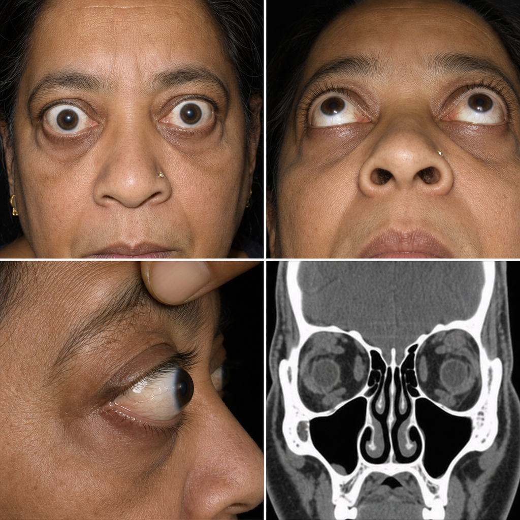
A patient with ptosis has the upper 4 mm of cornea covered by the upper eyelid. What is the grade of ptosis?
Xerophthalmia is considered a problem in a community if:
Which of the following is not included in the SAFE strategy?
Who is the chairman of the district blindness control society?
Explanation: ***All of the options*** - An ideal screening test must possess **all three essential attributes**: safety, reliability, and validity. - **Safe**: Minimizes harm to participants and ensures ethical implementation - **Reliable**: Produces consistent, reproducible results with minimal random error - **Valid**: Accurately measures what it intends to measure (high sensitivity and specificity) - These three attributes work together as fundamental requirements for any effective screening program, ensuring that early detection benefits outweigh potential risks. *Safe (alone)* - While safety is absolutely essential, it is **not sufficient by itself** to make an ideal screening test. - A test that is safe but unreliable or invalid would produce inconsistent or inaccurate results, rendering it ineffective for screening purposes. *Reliable (alone)* - Reliability ensures consistent results, which is crucial, but **reliability alone is insufficient**. - A test can be highly reliable (consistently giving the same result) yet completely invalid if it measures the wrong thing or is unsafe. *Valid (alone)* - Validity is critical for accurate measurement, but **validity alone does not make a test ideal**. - Even a valid test must be safe to protect participants and reliable to ensure consistency across different settings and times.
Explanation: ***Indirect ophthalmoscopy*** - While useful for viewing the peripheral retina, **indirect ophthalmoscopy** is less effective than other methods for assessing the optic nerve head and retinal nerve fiber layer changes characteristic of open-angle glaucoma. - Its primary utility is for detecting **retinal detachment** or other peripheral retinal pathologies, which are not directly diagnostic of glaucoma. *Tonometry* - **Tonometry** measures the intraocular pressure (IOP), a primary risk factor for open-angle glaucoma, and is essential for monitoring treatment effectiveness. - Elevated IOP is a key indicator, though not always present, and normal-tension glaucoma exists. *Direct ophthalmoscopy* - **Direct ophthalmoscopy** allows for visualization of the optic nerve head, enabling detection of characteristic glaucoma changes such as **cupping** and loss of the neuroretinal rim. - This method is crucial for assessing **optic nerve damage**, a hallmark of glaucoma. *Perimetry* - **Perimetry**, or visual field testing, assesses the functional impact of glaucoma by detecting **peripheral vision loss**. - This test identifies specific patterns of visual field defects that correlate with nerve fiber layer damage and is vital for staging and monitoring disease progression.
Explanation: ***Vision centre*** - Under Vision 2020 initiatives, a **Vision Centre** serves as the primary point of contact for basic ophthalmic services, including **visual acuity screening** and referral. - These centers are designed to be accessible in local communities, allowing teachers and other local caregivers to refer school children for initial checks and appropriate management. *Centre for excellence* - A **Centre for Excellence** typically refers to a highly specialized institution with advanced diagnostic and treatment capabilities, research facilities, and complex surgical procedures, which is **beyond the scope** of basic visual acuity checking and initial referral. - Such centers handle more **complex or rare conditions** and are not the first point of contact for routine school-based screening. *Training centre* - A **Training Centre** is primarily dedicated to educating and skilling healthcare professionals, not to providing direct patient care or screening services to the general public. - While essential for developing skilled personnel, it is **not the appropriate facility** for a teacher to refer a child for a visual acuity check. *Service centre* - The term **Service Centre** is too broad and can refer to various types of facilities that provide any kind of service, but it does not specifically denote a healthcare facility for ophthalmic care under the Vision 2020 program. - It lacks the **specific medical context** and structured role established for vision screening.
Explanation: ***A-4, B-1, C-3, D-2*** - **Morphine** is contraindicated in **head injury** as it can increase intracranial pressure and mask neurological symptoms. - **Amiodarone** is contraindicated in patients with **QT prolongation** due to its risk of inducing more severe arrhythmias like Torsades de Pointes. - **Vigabatrin** is contraindicated during **pregnancy** due to its potential for teratogenicity and adverse effects on fetal development. - **Estrogen preparations** are contraindicated in patients with a history of **thromboembolism** due to their increased risk of blood clot formation. *A-1, B-3, C-2, D-4* - This option incorrectly matches **Morphine** with QT prolongation and **Estrogen preparations** with head injury, which are not their primary contraindications. - It also incorrectly links **Vigabatrin** with thromboembolism and **Amiodarone** with pregnancy. *A-3, B-2, C-4, D-1* - This choice incorrectly associates **Morphine** with pregnancy and **Vigabatrin** with head injury, which are not the most critical or direct contraindications. - It also misaligns **Amiodarone** with thromboembolism and **Estrogen preparations** with QT prolongation. *A-2, B-4, C-1, D-3* - This option incorrectly matches **Morphine** with thromboembolism and **Amiodarone** with head injury, which are not their most significant contraindications. - It also incorrectly links **Vigabatrin** with QT prolongation and **Estrogen preparations** with pregnancy.
Explanation: ***Antecubital vein*** - Fluorescein angiography requires rapid delivery of the dye into the **systemic circulation** for quick visualization of retinal and choroidal vasculature. - The **antecubital vein** is a readily accessible, large superficial vein that allows for quick and efficient intravenous dye injection. *Popliteal vein* - The popliteal vein is located behind the **knee** and is not a standard or practical site for routine intravenous injections, especially when rapid delivery to the eye is needed. - Its location makes it less accessible and potentially more uncomfortable for the patient compared to an arm vein. *Femoral vein* - The femoral vein is a large, deep vein in the **groin**, typically reserved for central venous access or specific procedures due to the increased risk of complications like infection or hematoma. - It is not routinely used for peripheral intravenous injections such as fluorescein, where a more superficial and accessible vein is preferred. *Subclavian vein* - The subclavian vein is a **central vein** located under the clavicle, accessed via a more invasive procedure, usually for central venous catheters or hemodialysis access. - It carries higher risks compared to peripheral venipuncture and is not chosen for simple diagnostic dye injections like fluorescein.
Explanation: ***Correct: Statements 1 & 2 are correct, 2 is not explaining 1*** **Analysis of Statement 1:** - A 59-year-old patient with **flaccid bullae** and **suprabasal acantholytic split** on histopathology is the classic presentation of **Pemphigus vulgaris** - The flaccid (easily ruptured) nature of bullae distinguishes it from tense bullae seen in bullous pemphigoid - The suprabasal location of the split (just above the basal layer) with acantholysis (loss of cell-to-cell adhesion) is pathognomonic - **Statement 1 is CORRECT** ✓ **Analysis of Statement 2:** - The **"row of tombstones" or "tombstone appearance"** is indeed a diagnostic histopathological feature of Pemphigus vulgaris - This appearance results from basal keratinocytes remaining attached to the basement membrane while suprabasal cells separate due to acantholysis - The intact basal cells standing upright resemble a row of tombstones - **Statement 2 is CORRECT** ✓ **Does Statement 2 explain Statement 1?** - Statement 2 describes a **histopathological appearance** (tombstone pattern) that is a **consequence** of the suprabasal split - However, it does NOT explain the **underlying cause** of the flaccid bullae or the suprabasal split - The true explanation involves **IgG autoantibodies against desmoglein 3 (and desmoglein 1)**, which attack intercellular adhesion structures (desmosomes), causing **acantholysis** - Therefore, **Statement 2 does NOT explain Statement 1** ✗ *Incorrect: Statement 2 is the correct explanation for Statement 1* - While both statements describe features of Pemphigus vulgaris, the tombstone appearance is a descriptive finding, not an explanatory mechanism *Incorrect: Statements 1 and 2 are incorrect* - Both statements are medically accurate descriptions of Pemphigus vulgaris features *Incorrect: Statement 1 is incorrect* - Statement 1 correctly describes the cardinal clinical and histopathological features of Pemphigus vulgaris
Explanation: ***Moderate*** - **Moderate ptosis** is defined as **3-4 mm of lid drooping** below the normal position - In this case, the upper eyelid covers **4 mm of the cornea**, which falls into the moderate category - The lid margin is typically **at or slightly below the superior limbus** in moderate ptosis - This degree of ptosis is **functionally significant** and may warrant surgical correction *Mild* - **Mild ptosis** is defined as **2 mm or less** of lid drooping - The upper lid margin is **above the superior limbus** but below the normal position - This patient has 4 mm coverage, which **exceeds the mild category** *Severe* - **Severe ptosis** is defined as **5 mm or more** of lid drooping below the normal position - The upper lid typically **covers the pupillary axis significantly** and causes marked visual obstruction - This patient's 4 mm coverage **does not reach severe criteria** *Profound* - **"Profound"** is not a standard term in ptosis grading systems - The standard classification uses **mild, moderate, and severe** as the three grades - If used, it would refer to extreme cases where the lid almost completely covers the pupil
Explanation: **Explanation:** Vitamin A deficiency (VAD) is a major public health concern in developing nations. To determine if Xerophthalmia constitutes a "public health problem" in a community, the World Health Organization (WHO) has established specific prevalence criteria for children aged 6 months to 6 years. **Why Option A is Correct:** Night blindness (X1N) is the earliest clinical symptom of Vitamin A deficiency. According to WHO criteria, if the prevalence of night blindness in a community exceeds **1%**, it is considered a significant public health problem. This threshold is a high-yield fact for NEET-PG as it represents the primary screening indicator. **Analysis of Incorrect Options:** * **B. Bitot spots >1%:** This is incorrect because the WHO threshold for Bitot spots (X1B) is actually **>0.5%**. * **C. Corneal ulceration >0.05%:** This is incorrect because the threshold for active corneal lesions (X2, X3A, X3B) is **>0.01%**. * **D. Corneal scarring >0.5%:** This is incorrect because the threshold for Xerophthalmia-related corneal scars (XS) is **>0.05%**. **High-Yield Clinical Pearls for NEET-PG:** * **WHO Classification (Xerophthalmia):** * X1N: Night blindness * X1B: Bitot’s spots * X2: Conjunctival xerosis * X3A: Corneal xerosis * X3B: Keratomalacia (involving >1/3rd of the cornea) * XS: Corneal scar * XF: Xerophthalmic fundus * **Biochemical Criteria:** Serum retinol levels **<10 μg/dl (0.35 μmol/L)** in >5% of the population also signifies a public health problem. * **Prophylaxis:** Under the National Vitamin A Prophylaxis Program, the first dose (1 lakh IU) is given at 9 months (with Measles vaccine), followed by 2 lakh IU every 6 months until age 5 (Total 9 doses/17 lakh IU).
Explanation: The **SAFE strategy** is a comprehensive public health approach recommended by the World Health Organization (WHO) for the elimination of **Trachoma** (caused by *Chlamydia trachomatis*) as a public health problem. ### **Why "Surveillance" is the Correct Answer** While surveillance is a general epidemiological principle, it is **not** one of the four specific pillars of the SAFE acronym. The strategy focuses on active intervention and prevention rather than just monitoring. ### **Explanation of the SAFE Components (Incorrect Options)** The acronym **SAFE** stands for: * **S – Surgery:** To treat the blinding stage of the disease (**Trachomatous Trichiasis**). * **A – Antibiotics:** To treat the active infection and reduce the community reservoir. The drug of choice is a single dose of **Azithromycin** (20 mg/kg up to 1g) or Tetracycline eye ointment. * **F – Facial hygiene:** To reduce transmission from eye and nasal secretions, especially in children. * **E – Environmental modification:** Improving access to water and sanitation (e.g., latrine construction) to reduce the breeding of **Musca sorbens** (the eye-seeking fly that acts as a vector). ### **High-Yield Clinical Pearls for NEET-PG** * **Trachoma Classification:** The WHO uses the **FISTO** classification (Follicular, Intense, Scarring, Trichiasis, Opacity). * **Target:** The goal of the Global Elimination of Trachoma (GET2020) was to eliminate trachoma by 2020; India was declared free of "infective trachoma" in 2017. * **Vector:** *Musca sorbens* is the primary fly vector involved in transmission. * **Surgery Type:** The preferred surgical procedure for trichiasis is **Bilamellar Tarsal Rotation (BTR)**.
Explanation: ### Explanation The **District Blindness Control Society (DBCS)** is the decentralized implementation unit of the **National Programme for Control of Blindness and Visual Impairment (NPCBVI)**. **1. Why the District Collector is correct:** The District Collector (or District Magistrate) serves as the **Chairman** of the DBCS. This is a strategic administrative decision because blindness control requires inter-sectoral coordination between health, education, and social welfare departments. An administrative head ensures better resource mobilization, financial oversight, and accountability of the program at the district level. **2. Why the other options are incorrect:** * **Programme Manager:** Usually a senior eye surgeon or health official who handles the day-to-day operations and technical execution, but does not hold the chair. * **District Health Officer (CMO/CDMO):** Typically serves as the **Vice-Chairman** or Member Secretary. While they oversee the medical aspects, they report to the Collector for administrative approvals. * **District Eye Surgeon:** Acts as the **Member Secretary** or technical lead. They are responsible for the clinical quality of surgeries and organizing screening camps. **3. High-Yield Clinical Pearls for NEET-PG:** * **NPCBVI Goal:** To reduce the prevalence of blindness to **0.25%** by 2025 (Current prevalence is approx. 0.36% as per 2015-19 survey). * **Definition of Blindness (NPCBVI):** Visual acuity **<3/60** in the better eye with best possible correction. * **Funding:** The DBCS receives grants-in-aid directly from the State Health Society to ensure a "bottom-up" approach. * **Main Cause of Blindness in India:** Cataract (approx. 66%), followed by Refractive Errors.
Get full access to all questions, explanations, and performance tracking.
Start For Free