Which of the following is NOT a core component of the WHO's global STI control strategy?
Specificity of a diagnostic test is defined as:
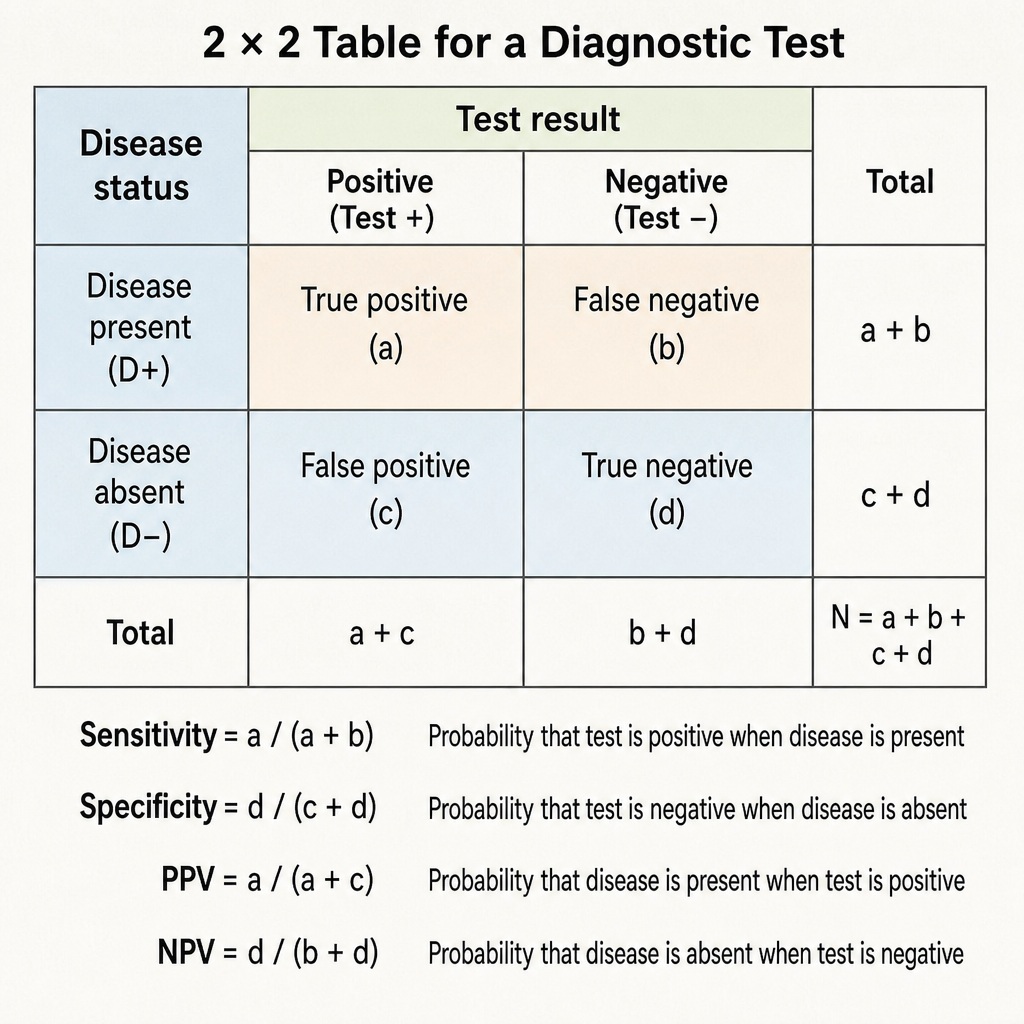
What is the correct formula for calculating the positive predictive value (PPV) of a screening test?
Under NPCB, screening of school children is first done by -
Which measure indicates the diagnostic power of a test to correctly identify those with a disease?
Which of the following procedures is not typically covered by the National Programme for Control of Blindness (NPCB) for reimbursement of surgery done by a non-governmental organization (NGO) eye hospital?
All of the following are the targets for National Programme for Control of Blindness (NPCB), EXCEPT:
Which of the following is a true statement about screening tests for genetic diseases?
In primary open-angle glaucoma (POAG), which of the following findings is NOT typically seen?
What is the gold standard method for visualizing the periphery of the retina?
Explanation: ***Universal mandatory screening*** - While screening is part of STI control, **universal mandatory screening** for all STIs in the general population is not a core component of the WHO's strategy due to feasibility, cost, and ethical considerations. - The strategy emphasizes **targeted screening** for at-risk populations and opportunistic screening. *Case management* - **Case management**, including accurate diagnosis and effective treatment, is a critical component for managing current infections and preventing further transmission. - This involves syndromic or etiologic approaches to treatment and partner notification. *Strategic information systems* - **Strategic information systems** are essential for monitoring trends, evaluating interventions, and informing policy decisions related to STI control. - This includes surveillance data, program monitoring, and research. *Prevention services* - **Prevention services** are a cornerstone of the WHO's strategy, aiming to reduce the incidence of new infections. - These services encompass health education, condom promotion and distribution, vaccination, and pre-exposure prophylaxis (PrEP).
Explanation: ***0.95*** - **Specificity** is the proportion of individuals without disease who test negative, calculated as **TN/(TN+FP)**. - A specificity of 0.95 (95%) indicates an excellent test that correctly identifies 95% of healthy individuals as negative. *0.05* - This value represents the **false positive rate** (1 - specificity), not specificity itself. - A specificity of 0.05 would mean only 5% of healthy individuals test negative, indicating a very poor test. *0.4* - This value is too low for specificity and could represent other test parameters like **positive predictive value**. - A specificity of 0.4 would incorrectly classify 60% of healthy individuals as positive, making the test clinically unreliable. *0.8* - This value typically represents **sensitivity**, which is the proportion of diseased individuals who test positive. - **Sensitivity** is calculated as **TP/(TP+FN)**, which is different from specificity that focuses on healthy individuals.
Explanation: ***True positives / (True positives + False positives)*** - **Positive predictive value (PPV)** indicates the probability that a patient who tests positive actually has the disease. - It is calculated by dividing the number of **true positives** (correctly identified positive cases) by the total number of positive test results (**true positives + false positives**). *True positives / (True positives + False negatives)* - This formula represents the **sensitivity** of a test, which is the proportion of actual positive cases that are correctly identified. - Sensitivity measures the ability of a test to correctly identify individuals with the disease. *False positives / (False positives + True negatives)* - This formula represents **1 - specificity**, or the **false positive rate**. - **Specificity** is the proportion of actual negative cases that are correctly identified as negative. *True negatives / (True negatives + False negatives)* - This formula represents the **negative predictive value (NPV)**, which is the probability that a patient who tests negative actually does not have the disease. - NPV is calculated by dividing the number of **true negatives** (correctly identified negative cases) by the total number of negative test results (**true negatives + false negatives**).
Explanation: ***School teachers*** - Under the **National Programme for Control of Blindness (NPCB)**, screening of school children follows a **three-tier approach**. - **School teachers** are trained to conduct the **first level/initial screening** using simple vision tests like **Snellen charts**. - They identify children with potential vision problems and refer them for further detailed assessment. - This approach maximizes coverage as teachers have regular contact with children and can screen large numbers efficiently. - The NPCB specifically includes **teacher training modules** for basic vision screening as part of the School Eye Screening Programme. *Ophthalmologic assistant* - Ophthalmic assistants/paramedical workers conduct the **second level screening** - the detailed assessment of children referred by teachers. - They perform comprehensive vision testing and identify specific refractive errors and eye conditions. - They are not the first point of contact in school screening due to resource limitations and the scale of screening required. *Medical officer* - Medical officers and ophthalmologists are involved in the **third tier** - providing diagnosis, treatment, and management of identified cases. - They handle complex cases, prescribe spectacles, and provide surgical interventions when needed. - They also supervise the overall program but do not conduct initial mass screening. *Village health guide* - Village health guides work primarily in community settings for general health promotion and basic healthcare. - While they contribute to community health awareness, they are not specifically involved in the structured school eye screening program under NPCB.
Explanation: ***Positive predictive value*** - It refers to the probability that subjects with a positive test result truly have the disease, highlighting the test's **diagnostic accuracy** [1]. - A high positive predictive value indicates that the test is effective at diagnosing the disease in the population tested. *Sensitivity* - Sensitivity measures the ability of a test to correctly identify those with the disease (true positives), but does not account for the test result's predictive capability [1]. - It is important for screening, but **not directly the diagnostic power** for those already tested. *Negative predictive value* - This indicates the probability that subjects with a negative test result truly do not have the disease, focusing on true negatives rather than correct diagnosis of the condition [1]. - While informative, it does not assess the ability to correctly diagnose the disease when the result is positive. *Specificity* - Specificity is the measure of a test's ability to correctly identify those without the disease (true negatives), not diagnosing the disease accurately among those tested [1]. - It is essential for determining false positives but not for assessing the overall diagnostic power of a test. **References:** [1] Cross SS. Underwood's Pathology: A Clinical Approach. 6th ed. (Basic Pathology) introduces the student to key general principles of pathology, both as a medical science and as a clinical activity with a vital role in patient care. Part 2 (Disease Mechanisms) provides fundamental knowledge about the cellular and molecular processes involved in diseases, providing the rationale for their treatment. Part 3 (Systematic Pathology) deals in detail with specific diseases, with emphasis on the clinically important aspects., pp. 253-254.
Explanation: ***Syringing and probing of the nasolacrimal duct*** - While important for lacrimal drainage issues, procedures like **syringing and probing** are generally considered minor and less vision-restoring compared to the major surgeries targeted by the **NPCB**. - The **NPCB** focuses on interventions for leading causes of blindness, primarily **cataract** and other significant vision-threatening conditions, which this procedure typically isn't. *Cataract surgery* - **Cataract surgery** is a cornerstone of the **NPCB's** efforts, as cataracts are the leading cause of reversible blindness. - Reimbursement for **cataract surgery** is a primary objective to improve access and reduce the burden of blindness. *Pan retinal photocoagulation for diabetic retinopathy* - **Diabetic retinopathy** is a major cause of preventable blindness, and **pan retinal photocoagulation (PRP)** is a key intervention to preserve vision. - The **NPCB** includes procedures for **diabetic retinopathy** management due to its significant public health impact. *Trabeculectomy surgery* - **Trabeculectomy** is a surgical procedure for **glaucoma**, which is another significant cause of irreversible blindness. - The **NPCB** includes interventions for **glaucoma** given its severe vision-threatening nature and the need for surgical management in many cases.
Explanation: ***100% coverage of vitamin A prophylaxis doses from 9 months to 3 years of age*** - This objective is typically associated with the **Reproductive and Child Health (RCH) program** and efforts to combat **Vitamin A deficiency**, not directly with the core targets of the National Programme for Control of Blindness (NPCB). - While vitamin A deficiency can lead to blindness, the NPCB's primary focus has historically been on treatable causes of blindness such as **cataract** and **refractive errors**. *Development of 50 pediatric ophthalmic units* - The NPCB aims to **strengthen ophthalmic services**, and the development of specialized pediatric units is a crucial component to address childhood blindness. - This target aligns with the program's focus on **preventing and treating blindness** across all age groups, including children. *Increase cataract surgery rate to 450 operations per lakh population* - **Cataract** is the leading cause of blindness in India, and increasing the **cataract surgery rate** is a key strategic objective of the NPCB to reduce the burden of preventable blindness. - This specific numerical target reflects the program's commitment to **scaling up surgical interventions**. *Intraocular lens implantation in more than 80 percent cataract surgery cases* - The NPCB emphasizes not just the quantity but also the **quality of cataract surgeries** performed. - Promoting **intraocular lens (IOL) implantation** in a high percentage of cases ensures better visual outcomes and rehabilitation for patients.
Explanation: ***Correct: It defines risk of transmission of disease to the child*** - Genetic screening aims to identify individuals or couples at risk of passing on **heritable genetic conditions** to their offspring. - This information helps in **family planning** and provides prenatal diagnostic options if the risk is high. - This is the **primary purpose** of genetic screening programs. *Incorrect: Screening test has better accuracy than diagnostic test* - **Screening tests** are designed to be broad and detect potential risks, often with lower specificity and sensitivity than diagnostic tests. - **Diagnostic tests** are typically more accurate and definitive, confirming the presence or absence of a disease after a positive screening result. *Incorrect: It is always invasive* - Many genetic screening tests, such as **non-invasive prenatal screening (NIPS)** from maternal blood or carrier screening via saliva, are non-invasive or minimally invasive. - While some diagnostic tests like **amniocentesis** or **chorionic villus sampling** are invasive, screening itself is not universally so. *Incorrect: Screening requires genetic mapping* - **Genetic mapping** refers to determining the relative positions of genes on a chromosome, which is a research tool for understanding genome organization. - Genetic screening primarily involves testing for specific mutations or chromosomal abnormalities, not creating a comprehensive genetic map of an individual.
Explanation: ***Macular edema*** - **Macular edema** is characterized by fluid accumulation in the fovea or retina, causing blurry vision and metamorphopsia. - While it can occur in conditions like **diabetic retinopathy** or **uveitis**, it is **not a typical finding in POAG**, which primarily affects the optic nerve. *Horizontal cupping of the optic disc* - **Horizontal cupping** (or vertical elongation of the optic cup) is a common sign of **glaucomatous damage**, reflecting the loss of retinal ganglion cells. - This morphological change suggests the progression of optic nerve atrophy. *Bayoneting sign (arteriovenous crossing changes)* - The **bayoneting sign**, where blood vessels appear to dip below the optic disc margin and then sharply bend, is a feature of **advanced glaucomatous cupping**. - It indicates significant loss of optic nerve tissue and is often associated with deeply excavated optic discs. *Peripapillary atrophy* - **Peripapillary atrophy** (PPA) refers to areas of RPE and choroidal atrophy surrounding the optic disc, commonly seen in glaucoma. - While its presence and extent may correlate with **glaucoma severity**, it is a recognized clinical feature of the disease.
Explanation: ***Correct: Indirect ophthalmoscopy*** - This method uses a **condensing lens** and a bright light source to provide a **wide-field, stereoscopic view** of the retina, making it ideal for visualizing the periphery. - It allows for examination even through some media opacities and is particularly useful for detecting peripheral retinal tears or detachments. - Provides a **field of view of 25-40 degrees** compared to only 5-10 degrees with direct ophthalmoscopy. *Incorrect: Direct ophthalmoscopy* - Provides a **highly magnified but narrow field of view**, making it difficult to systematically scan and visualize the entire peripheral retina. - It offers an **upright, monocular image** with limited depth perception, which is not optimal for assessing the three-dimensional structures of the retinal periphery. *Incorrect: Retinoscopy* - This is an objective method used to **determine the refractive error** of an eye, not for direct visualization of the retinal structures. - It involves observing the reflection of light from the retina as the examiner moves a light source across the eye. *Incorrect: USG* - **Ultrasound (USG)** is primarily used to visualize ocular structures when direct visualization is obscured by dense media opacities (e.g., severe cataracts, vitreous hemorrhage). - It provides 2D images and is not the gold standard for **routine, high-resolution visualization** of the retinal periphery when a clear view is obtainable.
Get full access to all questions, explanations, and performance tracking.
Start For Free