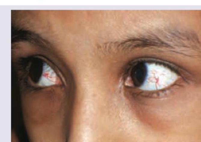
The image shows a child with?

Father of ophthalmology?
Which of the following is known as the dangerous zone of the eye?
Depth of anterior chamber (AC) is:
Which ocular structure is the most radiosensitive?
Which of the following statements about vitreous is false?
Which mode of ultrasonography is used to measure the axial length of the eyeball?
Muscae volitantes is seen in?
What is the normal aqueous production rate in the human eye?
Which of the following is a specific sign of albinism?
Explanation: ***Oculo-cutaneous albinism*** - The image exhibits signs consistent with **oculocutaneous albinism**, particularly the striking **transillumination defect** of the iris, where light passes through the iris, indicating a lack of melanin. - This condition is characterized by reduced or absent melanin production in the hair, skin, and eyes, leading to symptoms like **photophobia**, nystagmus, and reduced visual acuity. *Telangiectasia* - **Telangiectasia** refers to dilated small blood vessels visible on the skin or mucous membranes, typically appearing as fine red lines. - These are not the primary feature shown in the image; the prominent finding is the lack of iris pigmentation and visible fundus reflex through the iris. *Bitot's spots* - **Bitot's spots** are foamy, triangular patches of keratinized conjunctiva, often seen in the interpalpebral fissure, indicative of **Vitamin A deficiency**. - No such foamy patches are visible in the provided image. *Pterygium* - A **pterygium** is a benign growth of the conjunctiva that extends onto the cornea, typically wedge-shaped and often associated with UV exposure. - The image does not show any corneal encroachment by conjunctival tissue; rather, the underlying issue is related to pigmentation.
Explanation: ***Albrecht Von Graefe*** - **Albrecht von Graefe** is widely regarded as the **Father of Modern Ophthalmology** due to his pioneering contributions to the field. - He made significant advancements in understanding and treating **glaucoma**, **cataract surgery**, and **retinal detachment**. - His contributions revolutionized the surgical and clinical management of eye diseases in the 19th century. *William Osler* - **William Osler** is considered one of the **Fathers of Modern Medicine**, particularly known for his contributions to medical education and clinical practice. - His work focused on broad internal medicine, not specifically ophthalmology. *Sushruta* - **Sushruta** was an ancient Indian physician who is often called the **Father of Surgery** for his detailed descriptions of surgical procedures. - While his work included some eye-related surgeries (particularly cataract surgery), he is not specifically recognized as the father of modern ophthalmology. *Hermann von Helmholtz* - **Hermann von Helmholtz** was a German physician and physicist renowned for his contributions to optics and physiology. - He **invented the ophthalmoscope** in 1851, a crucial tool for examining the eye, but is not considered the overall father of the entire field of ophthalmology.
Explanation: ***Ciliary body*** - The **dangerous zone** (or dangerous area) of the eye refers to the region approximately **3-4 mm posterior to the limbus**, which overlies the **pars plana of the ciliary body**. - This area is termed "dangerous" because penetrating injuries or surgical trauma in this zone can result in multiple serious complications: - **Ciliary body damage** → Hypotony, hemorrhage, sympathetic ophthalmia - **Lens injury** → Traumatic cataract formation - **Vitreous involvement** → Endophthalmitis, vitreous hemorrhage, retinal detachment - This zone is clinically significant because the **extraocular muscles insert** near this region, and it represents the thinnest part of the sclera with underlying vital structures. *Sclera* - While the sclera forms the outer protective coat of the eye and can be vulnerable to trauma, the term "dangerous zone" specifically refers to a particular region (overlying the ciliary body), not the sclera as a whole. - The sclera provides structural support but is not itself called the dangerous zone. *Optic nerve* - The optic nerve transmits visual information from the retina to the brain and damage causes irreversible vision loss. - However, it is not referred to as the "dangerous zone" in ophthalmological terminology. *Retina* - The retina is the light-sensitive neurosensory tissue essential for vision. - Retinal damage leads to vision loss (e.g., retinal detachment, macular degeneration), but it is not termed the "dangerous zone."
Explanation: ***2-3 mm*** - The **anterior chamber (AC)**, the space between the cornea and the iris/lens, typically has a depth of **2.5-3.0 mm** (commonly cited as **2-3 mm**) in a healthy adult eye. - This measurement is important for assessing ocular health, particularly in conditions like **glaucoma** or before intraocular surgery. *1-2 mm* - A depth of **1-2 mm** would be considered **shallow**, often indicative of a **narrow angle** and increased risk for conditions like **acute angle-closure glaucoma**. - This could also be seen in individuals with **hyperopia** or certain anatomical variations. *4-5 mm* - An anterior chamber depth of **4-5 mm** is considered **deep** and exceeds the normal range for most adults. - While it may be seen in certain conditions like **high myopia**, **buphthalmos**, or after specific surgical interventions, it is not within the typical normal range. *6-8 mm* - An anterior chamber depth of **6-8 mm** is considered unusually **deep** and is rare for a healthy adult eye. - This degree of deepening might be observed in severe pathological conditions or after specific surgical interventions.
Explanation: ***Lens*** - The **lens** is the most radiosensitive structure of the eye, particularly its **equatorial epithelial cells**. - Radiation exposure can lead to the development of **cataracts**, a common long-term complication in individuals exposed to ionizing radiation. *Cornea* - The **cornea** is relatively radioresistant compared to the lens. - While high doses of radiation can cause corneal damage such as **keratitis**, it is less susceptible to radiation-induced pathology at doses that affect the lens. *Retina* - The **retina** is also relatively radioresistant compared to the lens, although very high doses can cause **retinopathy**. - Symptoms of radiation retinopathy include **microaneurysms**, hemorrhages, and eventually vision loss, but these require significantly higher doses than those causing cataracts. *All of the options* - This option is incorrect because while all mentioned structures can be affected by radiation, they exhibit different sensitivities. - The **lens** stands out as uniquely vulnerable, making it the most radiosensitive among them.
Explanation: ***Anatomically, vitreous is present in anterior segment*** - The vitreous humor is located in the **posterior segment** of the eye, filling the space between the lens and the retina. - The **anterior segment** comprises structures anterior to the vitreous, such as the cornea, iris, ciliary body, and lens. *Vitreous largely contains water and hyaluronic acid* - The vitreous body is approximately **99% water**, with the remaining 1% consisting mainly of **collagen fibrils** and **hyaluronic acid**. - Hyaluronic acid contributes significantly to the **viscoelastic properties** and gel-like consistency of the vitreous. *Strongest attachment of vitreous base is at ora serrata* - The **vitreous base** is the strongest area of attachment of the vitreous to the retina and is located circumferentially at the **ora serrata**. - This strong bond is crucial for maintaining the position of the vitreous and has implications in conditions like **retinal tears**. *Vitreous is attached anteriorly to the lens* - The anterior surface of the vitreous, known as the **anterior hyaloid membrane**, is attached to the posterior capsule of the lens in a ring-like fashion called **Wieger's ligament**. - This attachment is typically firm in younger individuals but can weaken with age, leading to conditions like **posterior vitreous detachment**.
Explanation: ***A-mode Ultrasonography*** - **A-mode** (amplitude modulation) ultrasonography is a **one-dimensional** display that measures the distance between structures based on the time it takes for sound waves to reflect. - It is specifically used for **biometry**, such as measuring the axial length of the eyeball for **intraocular lens (IOL) power calculation** prior to cataract surgery. *B-mode Ultrasonography* - **B-mode** (brightness modulation) ultrasonography provides a **two-dimensional** image, displaying the cross-sectional anatomy of organs. - While useful for visualizing ocular structures, it is not primarily used for precise **axial length measurements**. *M-mode Ultrasonography* - **M-mode** (motion mode) ultrasonography displays the **movement** of structures over time in a one-dimensional format. - This mode is typically used in **cardiac imaging** to assess heart valve motion and chamber dimensions, not for static length measurements of the eye. *None of the options* - This option is incorrect because **A-mode ultrasonography** is indeed the gold standard for measuring the axial length of the eyeball. - The other modes (B-mode and M-mode) serve different diagnostic purposes in ultrasonography.
Explanation: ***Vitreous detachment*** - **Muscae volitantes** (Latin for "flying flies") is the classical term for **vitreous floaters** - **Posterior vitreous detachment (PVD)** is the **most common cause** of muscae volitantes - PVD occurs due to **vitreous liquefaction** and **vitreoretinal separation**, causing **collagen fibers and cellular debris** to cast shadows on the retina - These floaters are typically described as **spots, cobwebs, or threads** that move with eye movement - PVD is extremely common, particularly **after age 50-60 years**, and in **myopic patients** *Remains of primitive hyaloid vasculature* - **Hyaloid remnants** (such as **Mittendorf's dot** or **Bergmeister's papilla**) are **congenital developmental anomalies** - While these can occasionally be visible as small opacities, they are **not the classical cause** of muscae volitantes - These are typically **stationary** rather than "floating" as the term implies *Vitreous Hemorrhage* - Causes **sudden onset** of numerous floaters, often described as **"shower of red blood cells"** or dense black spots - Associated with **significant vision loss** and requires urgent evaluation - Not the benign, chronic floaters typical of muscae volitantes *Eale's disease* - **Peripheral retinal vasculitis** causing **recurrent vitreous hemorrhages** - Floaters occur secondary to **hemorrhage**, not the primary pathology of muscae volitantes - Presents with **recurrent episodes** and **vision loss**, not isolated benign floaters
Explanation: ***2 µl/min*** - The ciliary body in the human eye continuously produces **aqueous humor** at a typical rate of approximately **2.0-3.0 µl/min**. - This rate is crucial for maintaining **intraocular pressure (IOP)** and providing nutrients to avascular eye structures like the **cornea** and **lens**. - This translates to approximately **3-4 ml per day** of aqueous humor production. *2 ml/min* - This rate is significantly **higher** than the actual production of aqueous humor, which is measured in microliters per minute. - Such a high production rate would lead to a rapid and severe increase in **intraocular pressure**, causing damage to the eye. *5 ml/min* - This value is an **excessively high** rate of fluid production and is not physiologically correct for aqueous humor. - It would result in unsustainable and damaging **intraocular pressure** levels. *5 µl/min* - While closer to the correct order of magnitude, **5 µl/min** is generally considered to be at the higher end or slightly above the average physiological range for aqueous humor production. - Most sources cite the normal range as being between **2-3 µl/min**.
Explanation: ***Iris transillumination*** - This is a highly **specific sign** of albinism, resulting from the severe reduction or absence of pigment in the iris. - When light shines through the pupil, it passes through the unpigmented iris, creating a visible red reflex, indicating the lack of pigment that normally blocks the light. *Sensitivity to light (photophobia)* - While common in albinism due to the lack of pigment in the iris and retina allowing more light to enter the eye, **photophobia is not specific** to albinism. - It can be a symptom of various other ocular conditions like uveitis, corneal abrasions, or migraines. *Involuntary eye movements (nystagmus)* - **Nystagmus is frequently associated with albinism** due to foveal hypoplasia and impaired visual development but is **not specific**. - It can also be caused by neurological disorders, inner ear problems, or other ocular conditions. *Decreased visual acuity* - **Reduced vision is a characteristic feature of albinism** resulting from foveal hypoplasia and abnormal optic nerve pathways, but it is **not specific** to the condition. - Numerous eye conditions, such as refractive errors, cataracts, and retinal diseases, can lead to decreased visual acuity.
Embryology of Eye
Practice Questions
Ocular Anatomy
Practice Questions
Ocular Physiology
Practice Questions
Ocular Biochemistry
Practice Questions
Ocular Microbiology
Practice Questions
Ocular Pharmacology
Practice Questions
Ocular Pathology
Practice Questions
Ocular Genetics
Practice Questions
Ocular Immunology
Practice Questions
Visual Neuroscience
Practice Questions
Ocular Imaging Physics
Practice Questions
Laser Physics in Ophthalmology
Practice Questions
Get full access to all questions, explanations, and performance tracking.
Start For Free